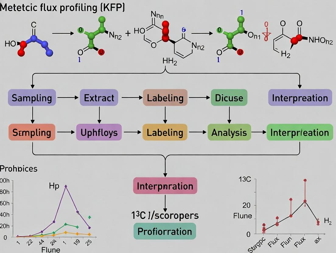
Unraveling Metabolic Pathways: A Comprehensive Guide to 13C Kinetic Flux Profiling (KFP) Protocol
This article provides a detailed, step-by-step guide to the 13C Kinetic Flux Profiling (KFP) protocol for researchers and drug development professionals.
Unraveling Metabolic Pathways: A Comprehensive Guide to 13C Kinetic Flux Profiling (KFP) Protocol
Abstract
This article provides a detailed, step-by-step guide to the 13C Kinetic Flux Profiling (KFP) protocol for researchers and drug development professionals. Covering foundational principles, methodological execution, and advanced applications, it explores how KFP quantifies intracellular metabolic fluxes using stable isotope tracers. We detail experimental design, data acquisition via mass spectrometry, computational flux analysis, and troubleshooting common pitfalls. The content validates KFP against other flux analysis methods and highlights its crucial role in identifying metabolic vulnerabilities in disease and for evaluating drug mechanisms of action in preclinical research.
What is 13C Kinetic Flux Profiling? Core Principles and Scientific Rationale
1. Introduction: Thesis Context and Rationale
This application note is framed within a broader thesis research program focused on advancing the 13C Kinetic Flux Profiling (KFP) protocol. While classical steady-state 13C Metabolic Flux Analysis (MFA) provides a snapshot of net fluxes through metabolic networks at isotopic equilibrium, it lacks temporal resolution for dynamic processes. KFP addresses this by quantifying the time-dependent labeling of metabolites following the introduction of a 13C tracer, thereby enabling the determination of absolute intracellular flux rates (in µmol/gDW/min) and pool sizes. This protocol is critical for research in systems biology, cancer metabolism, and drug development, where understanding metabolic adaptation and target engagement is paramount.
2. Core Principles of Kinetic Flux Profiling
KFP utilizes dynamic 13C labeling data, typically from LC-MS measurements, to fit parameters of an ordinary differential equation (ODE) model representing the metabolic network. The fitted parameters are the unidirectional fluxes (V) and metabolite pool sizes (Q). This contrasts with steady-state MFA, which solves for net fluxes at isotopic steady-state. KFP's requirement for precise time-series data and sophisticated computational fitting presents both a challenge and a source of richer biological insight.
3. Application Notes: Key Insights from Recent Studies
Recent applications of KFP have elucidated rapid metabolic rewiring in response to stimuli. The following table summarizes quantitative findings from key studies illustrative of the KFP approach.
Table 1: Summary of Quantitative Insights from Recent KFP Studies
| Biological System | Perturbation | Key Metabolic Finding via KFP | Quantified Change (Example) | Implication |
|---|---|---|---|---|
| Cultured Cancer Cells | Acute EGF stimulation | Glycolytic flux increase precedes TCA cycle change | VPFK increased by 80% within 2 minutes | Signaling-driven metabolic prioritization |
| Activated T Cells | Immune receptor engagement | Anaplerotic pyruvate carboxylase (PC) flux surge | VPC increased 5-fold within 1 hour | Supports biomass for proliferation |
| Hepatocytes | Glucagon exposure | Rapid diversion of gluconeogenic flux | VPEPCK doubled within 10 minutes | Hormonal control of metabolic routing |
| Drug-Treated Cells (Thesis Focus) | OXPHOS inhibitor (e.g., Metformin) | Compensatory glycolysis and serine biosynthesis flux | VPHGDH increased by 150% | Identifies potential drug resistance pathways |
4. Detailed Experimental Protocol: 13C-KFP in Mammalian Cells
4.1. Materials and Reagent Solutions
Table 2: The Scientist's Toolkit - Key Reagents for 13C-KFP
| Reagent / Material | Function / Explanation |
|---|---|
| U-13C-Glucose (or other tracer) | Uniformly labeled substrate to initiate labeling kinetics; defines the entry point of label. |
| Custom, Serum-Free Labeling Medium | Chemically defined medium necessary for precise control of extracellular nutrient concentrations. |
| Rapid Sampling Apparatus (e.g., Vacuum Filtration) | Enables quenching of metabolism and collection of samples at sub-second to minute intervals. |
| Pre-chilled Quenching Solution (e.g., 60% Methanol -40°C) | Instantly halts enzymatic activity to preserve metabolic state at time of sampling. |
| LC-MS/MS System with High Resolution | For accurate quantification of metabolite concentrations (via unlabeled peaks) and 13C isotopologue distributions. |
| Computational Software (e.g., INCA, Q-Flux) | Used for kinetic model construction, experimental data integration, and non-linear parameter fitting. |
4.2. Step-by-Step Protocol
Day 1: Cell Preparation
- Seed cells in appropriate dishes to reach 70-80% confluence at time of experiment.
- 24 hours prior to experiment, switch cells to custom serum-free, chemically defined medium matching the future labeling medium except for the tracer.
Day 2: Kinetic Labeling Experiment
- Pre-equilibration: Aspirate medium and wash cells twice with pre-warmed PBS. Add pre-warmed labeling medium containing natural abundance nutrients. Incubate for 60 min to establish a metabolic steady-state under experimental conditions.
- Tracer Switch & Rapid Sampling: Rapidly aspirate medium and immediately add labeling medium containing the 13C tracer (e.g., [U-13C] Glucose). Start timer.
- Sample cells at a pre-determined time series (e.g., 0, 15s, 30s, 1m, 2m, 5m, 10m, 20m, 30m, 60m).
- For adherent cells: Use rapid vacuum aspiration/quenching with cold methanol or direct scraping into quenching solvent.
- Sample Processing: Extracellular medium is also collected at each time point for exometabolite analysis. Cell pellets are extracted using a methanol/water/chloroform extraction. Extracts are centrifuged, and the aqueous phase is dried and stored at -80°C for LC-MS analysis.
Day 3-4: LC-MS Metabolomics
- Reconstitute samples in MS-compatible solvent.
- Run samples on a hydrophilic interaction liquid chromatography (HILIC) or ion-pairing LC system coupled to a high-resolution mass spectrometer.
- Acquire data in full-scan mode to quantify isotopologue distributions (M+0, M+1, M+2, … M+n) for target metabolites (e.g., glycolytic intermediates, TCA cycle acids, amino acids).
- Quantify absolute pool sizes using internal standards and calibration curves run in parallel.
Day 5-7: Computational Modeling & Flux Estimation
- Model Construction: Define a stoichiometric network model including atom transitions.
- Data Integration: Input the measured time-course data: extracellular nutrient/ byproduct rates, intracellular pool sizes (from M+0), and isotopologue fractions.
- Parameter Fitting: Use non-linear least squares optimization to fit the unknown parameters (V, Q) to the labeling kinetics data. This step minimizes the difference between model-simulated and experimentally measured isotopologue time courses.
- Statistical Analysis: Perform Monte Carlo simulations or sensitivity analysis to estimate confidence intervals for the fitted fluxes and pool sizes.
5. Visualizing the KFP Workflow and Metabolic Network
13C Kinetic Flux Profiling (KFP) Core Workflow
Simplified Network for 13C-KFP of Central Metabolism
The Central Role of 13C-Labeled Tracers in Dynamic Flux Measurement
Kinetic Flux Profiling (KFP) is a cornerstone methodology within metabolic research, enabling the quantitative, time-resolved measurement of intracellular reaction rates (fluxes). At the heart of KFP is the use of 13C-labeled tracers, which provide the temporal dimension necessary to observe pathway dynamics, rather than static snapshots. This application note details the protocols and critical considerations for implementing 13C-based KFP within a drug discovery and biomedical research context, where understanding metabolic rewiring is essential.
13C-KFP tracks the incorporation of stable isotope atoms from a labeled nutrient (e.g., [U-13C]glucose) into downstream metabolites over time. The resulting labeling patterns and kinetics are used with computational models to infer absolute metabolic fluxes.
Table 1: Common 13C-Labeled Tracers and Their Applications in KFP
| Tracer Compound | Labeling Pattern | Primary Pathway Interrogated | Typical Concentration Range | Key Measured Fluxes |
|---|---|---|---|---|
| Glucose | [U-13C] | Glycolysis, PPP, TCA Cycle | 5-25 mM (match media) | Glycolytic flux, Pyruvate dehydrogenase/ carboxylase flux |
| Glucose | [1-13C] | Pentose Phosphate Pathway (PPP) | 5-25 mM | Oxidative vs. non-oxidative PPP flux |
| Glutamine | [U-13C] | Anaplerosis, TCA Cycle, Reductive carboxylation | 2-6 mM (match media) | Glutaminolysis flux, α-KG dehydrogenase flux |
| Acetate | [U-13C] | Acetyl-CoA metabolism | 1-5 mM | Cytosolic vs. mitochondrial acetyl-CoA usage |
| 13C-Lactate | [3-13C] | Gluconeogenesis, Cori cycle | 1-10 mM | Lactate uptake, PC flux |
Table 2: Mass Spectrometry Platforms for 13C-KFP Analysis
| Platform Type | Measured Ions | Typical Time Resolution (for KFP) | Key Advantage for KFP |
|---|---|---|---|
| GC-MS (Quadrupole) | Fragmentation patterns of derivatized metabolites | 5-15 minutes | High reproducibility, extensive libraries |
| LC-MS (Q-TOF/Orbitrap) | Intact metabolite masses (M+, M+1, M+2... M+n) | 2-10 minutes | Broad coverage without derivatization, high mass accuracy |
| LC-MS/MS (TQMS) | Specific fragment ions | 3-7 minutes | Superior sensitivity for low-abundance metabolites |
Detailed Experimental Protocols
Protocol 1: Cell Culture Pulse-Chase Experiment for Central Carbon Metabolism KFP
Objective: To determine dynamic fluxes in glycolysis, TCA cycle, and associated pathways.
Materials:
- Adherent or suspension cells (e.g., cancer cell line, primary hepatocytes)
- Standard growth medium (e.g., DMEM, RPMI)
- Custom tracer medium: Identical composition but with natural glucose replaced by [U-13C]glucose.
- Phosphate-Buffered Saline (PBS), pre-warmed and isotope-free.
- Quenching solution: 60% methanol (LC-MS grade) in water, chilled to -40°C to -80°C.
- Extraction solvent: 80% methanol/water at -80°C.
- Cell scraper or equivalent.
- Centrifuge and vortexer.
- SpeedVac concentrator.
- GC-MS or LC-MS autosampler vials.
Procedure:
- Culture & Preparation: Grow cells to desired confluence (typically 70-80%) in standard medium. Use biological replicates (n≥3).
- Medium Exchange: Rapidly aspirate standard medium. Wash cells twice quickly with pre-warmed PBS to remove natural nutrient traces.
- Pulse Initiation (t=0): Immediately add pre-warmed tracer medium containing [U-13C]glucose.
- Time-Course Sampling: At defined time points (e.g., 0, 15s, 30s, 1min, 2min, 5min, 10min, 30min, 60min): a. Rapidly aspirate tracer medium. b. Immediately add chilled quenching solution (e.g., 1 mL for a 6-well plate well). Place plate on dry ice or at -80°C.
- Metabolite Extraction: Scrape cells in the quenching solution. Transfer cell suspension to a pre-chilled microcentrifuge tube. a. Vortex vigorously for 30 seconds. b. Centrifuge at max speed (>15,000 g) for 10 minutes at 4°C. c. Transfer supernatant to a new tube. d. Dry the supernatant in a SpeedVac concentrator (no heat).
- Sample Analysis: Resuspend dried metabolites in appropriate solvent for MS analysis (e.g., water for LC-MS, methoxyamine/pyridine for GC-MS). Analyze via GC-MS or LC-MS.
Protocol 2: LC-MS/MS Method for Key 13C-Labeled Metabolite Quantification
Objective: To separate and detect the mass isotopomer distribution (MID) of central carbon metabolites.
Chromatography (HILIC Method Example):
- Column: SeQuant ZIC-pHILIC (5 µm, 2.1 x 150 mm)
- Mobile Phase A: 20 mM ammonium carbonate, 0.1% ammonium hydroxide in water
- Mobile Phase B: Acetonitrile
- Gradient: 80% B to 20% B over 20 min, hold 5 min, re-equilibrate.
- Flow Rate: 0.15 mL/min
- Column Temperature: 40°C
- Injection Volume: 5-10 µL
Mass Spectrometry (Q-TOF Example):
- Ionization: Electrospray Ionization (ESI), negative mode for organic acids, positive for amino acids.
- Scan Range: m/z 70-1000
- Source Conditions: Gas Temp 250°C, Drying Gas 10 L/min, Nebulizer 30 psi.
- Data Acquisition: Full scan mode for accurate mass. Data-dependent MS/MS for verification.
- Data Analysis: Use software (e.g., Agilent MassHunter, XCMS, or custom MATLAB/Python scripts) to extract chromatographic peaks and calculate MIDs for metabolites of interest (e.g., glucose-6P, lactate, citrate, malate, aspartate).
Visualization of KFP Concepts and Workflows
Title: KFP Experimental and Computational Workflow
Title: 13C Flow from Glucose to Early TCA Cycle
The Scientist's Toolkit: Essential Reagents and Materials
Table 3: Key Research Reagent Solutions for 13C-KFP
| Item | Function/Benefit in KFP | Example Product/Catalog Number (Informational) |
|---|---|---|
| [U-13C]Glucose | Uniformly labeled tracer for comprehensive mapping of carbon fate through glycolysis, PPP, and TCA cycle. Essential for mass balance. | CLM-1396 (Cambridge Isotope Labs) |
| [U-13C]Glutamine | Critical tracer for analyzing glutaminolysis, anaplerotic flux into TCA cycle, and reductive carboxylation in conditions like hypoxia. | CLM-1822 (Cambridge Isotope Labs) |
| Isotope-Free Base Medium | Custom formulated medium lacking the target nutrient (e.g., glucose- or glutamine-free DMEM) for precise tracer medium preparation. | Various (e.g., US Biological, Sigma) |
| Pre-chilled 60% Methanol | Quenching solution that rapidly halts metabolic activity, preserving the in vivo labeling state at the moment of sampling. | Prepared in-house (LC-MS grade) |
| Derivatization Reagent (for GC-MS) | Converts polar metabolites to volatile derivatives (e.g., MOX-TBDMS). Enables gas chromatography separation. | N-Methyl-N-(tert-butyldimethylsilyl)trifluoroacetamide (MTBSTFA) |
| HILIC Chromatography Column | Separates highly polar, water-soluble metabolites (sugar phosphates, organic acids) for LC-MS analysis. | SeQuant ZIC-pHILIC (MilliporeSigma) |
| Internal Standard Mix (13C or 15N labeled) | Corrects for variation in extraction efficiency and instrument response during MS analysis. | MSK-CUS-INDY (Cambridge Isotope Labs) |
| Flux Analysis Software | Performs computational modeling to convert time-course MID data into quantitative flux maps. | INCA, IsoCor, OpenFLUX, 13C-FLUX2 |
Application Notes
13C Kinetic Flux Profiling (KFP) has emerged as a pivotal methodology for quantifying metabolic flux dynamics in living cells. Within the broader thesis on KFP protocol research, this approach directly addresses the core biological question of how intracellular pathway activity is reprogrammed in response to therapeutic intervention, thereby predicting and explaining drug response. Recent advances have demonstrated its utility from basic biology to translational drug development.
Connecting Flux to Phenotype: A primary application is the quantification of flux rewiring in cancer models upon treatment with targeted therapies (e.g., kinase inhibitors) or chemotherapies. KFP can reveal compensatory metabolic pathways that enable cell survival, identifying potential drug targets for combination therapies. For instance, increased glutaminase flux is a known resistance mechanism to PI3K/mTOR inhibitors.
Pharmacodynamic Assessment: KFP serves as a powerful pharmacodynamic (PD) biomarker tool. By tracing 13C-labeled nutrients (e.g., [U-13C]-glucose, [U-13C]-glutamine) into downstream metabolites, researchers can measure the in vivo modulation of specific pathway activities (like glycolysis, TCA cycle, or pentose phosphate pathway) within hours of drug administration, far earlier than tumor volume changes.
Predictive Biomarker Discovery: Pre-treatment fluxomic profiles can classify tumors based on their metabolic dependencies. Tumors reliant on oxidative phosphorylation (OxPhos) may be intrinsically resistant to glycolytic inhibitors but sensitive to mitochondrial poisons. KFP enables the functional annotation of these states beyond genomic signatures.
Quantitative Data Summary:
Table 1: Representative KFP-Derived Flux Changes in Cancer Cell Lines Upon Drug Treatment
| Drug Class (Example) | Target Pathway | Key Flux Alteration (Measured by KFP) | Fold-Change Range | Implication for Response |
|---|---|---|---|---|
| PI3K/mTOR Inhibitor (e.g., Pictilisib) | Glycolysis, PPP | ↓ Glycolytic flux to lactate; ↑ OxPhos; ↑ Pentose Phosphate Pathway flux | Glycolysis: 0.3-0.7x; PPP: 1.5-3.0x | Compensatory NADPH production; Resistance via metabolic plasticity |
| IDH1 Inhibitor (e.g., Ivosidenib) | TCA Cycle | ↓ D-2-hydroxyglutarate production; ↑ glutaminolysis | D2HG: <0.1x; Gln Anaplerosis: 1.8-2.5x | On-target efficacy; Possible adaptive fueling |
| Chemotherapy (e.g., Doxorubicin) | Nucleotide Synthesis | ↑ Pyrimidine de novo synthesis flux from glucose | 2.0-4.0x | Increased demand for DNA repair; Target for sensitization |
| Glutaminase Inhibitor (e.g., CB-839) | Amino Acid Metabolism | ↓ Malate from glutamine; ↑ glucose-derived anaplerosis | Gln→Malate: 0.2-0.5x | Efficacy in glutamine-addicted models; Resistance via glucose fueling |
Table 2: Essential 13C-Labeled Tracers for Drug Response Studies
| Tracer | Primary Pathways Probed | Typical Concentration | Key Drug Response Questions |
|---|---|---|---|
| [U-13C]-Glucose | Glycolysis, PPP, TCA Cycle, Serine Synthesis | 5-25 mM (culture media) | How does drug X affect glycolytic commitment vs. mitochondrial oxidation? |
| [U-13C]-Glutamine | Glutaminolysis, TCA Cycle (anaplerosis), Redox balance | 2-4 mM | Does the drug impair glutamine-fueled biomass/energy production? |
| [1,2-13C]-Glucose | PPP vs. Glycolysis partitioning | 5-25 mM | Is the oxidative PPP induced as a survival mechanism? |
| [U-13C]-Palmitate (with BSA) | Fatty Acid Oxidation (FAO) | 100-200 µM | Does therapy induce a dependency on mitochondrial FAO? |
Detailed Protocols
Protocol 1: KFP Workflow forIn VitroDrug Response Profiling
Objective: To quantify acute changes in central carbon metabolism flux following drug treatment in adherent cancer cell lines.
I. Materials & Cell Preparation
- Cells in mid-log phase.
- Drug of interest and vehicle control.
- Customized tracer media: DMEM-based, lacking glucose and glutamine, supplemented with dialyzed FBS, 10 mM [U-13C]-Glucose, and 4 mM [U-13C]-Glutamine.
- Quenching solution: 60% cold aqueous methanol (-40°C).
- LC-MS system with appropriate columns (e.g., HILIC for polar metabolites).
II. Procedure
- Seed & Treat: Seed cells at 70% confluence. After 24h, treat with drug or vehicle for the desired pharmacodynamic window (e.g., 2, 6, 24h).
- Tracer Pulse: At the end of drug treatment, quickly aspirate media and replace with pre-warmed 13C tracer media. Incubate for a precisely timed pulse (typically 15 min to 2 h, optimized for linear incorporation).
- Rapid Metabolite Extraction:
- Aspirate tracer media and immediately wash plates with 5 mL of ice-cold 0.9% NaCl.
- Add 1 mL of quenching solution, then scrape cells on dry ice.
- Transfer extract to a cold tube, vortex, and incubate at -40°C for 30 min.
- Centrifuge at 16,000 x g for 15 min at -9°C. Transfer supernatant to a new tube.
- Dry under a gentle stream of nitrogen or in a speed vacuum.
- LC-MS Sample Prep & Analysis:
- Reconstitute dried extracts in 100 µL of LC-MS compatible solvent (e.g., water:acetonitrile, 1:1).
- Centrifuge and transfer to MS vials.
- Run on a HILIC column coupled to a high-resolution mass spectrometer.
- Acquire data in negative/positive ion switching mode to cover a broad metabolome.
- Data Processing: Use software (e.g., El-MAVEN, XCMS) to integrate peaks. Correct for natural isotope abundance and calculate 13C isotopologue distributions (MIDs) for key metabolites.
Protocol 2:In VivoFlux Analysis of Tumor Drug Response
Objective: To measure tumor metabolic flux in situ following drug administration in a mouse xenograft model.
I. Materials
- Tumor-bearing mice (subcutaneous or orthotopic).
- Sterile [U-13C]-Glucose solution in PBS (e.g., 1.5 g/kg body weight).
- Infusion pump or materials for bolus injection.
- Drug or vehicle control.
- Liquid nitrogen or clamp freezer for tissue fixation.
II. Procedure
- Drug Treatment: Administer drug or vehicle to mice at therapeutically relevant dose and schedule.
- 13C-Glucose Infusion: At the pharmacodynamic peak (e.g., 2h post-drug), anesthetize mouse. Perform a tail-vein bolus injection or start a primed, constant infusion of [U-13C]-glucose over a defined period (e.g., 10-30 min).
- Rapid Tissue Collection: At the end of infusion, euthanize the animal and immediately excise the tumor. Submerge tissue in liquid nitrogen within 5-10 seconds. Store at -80°C.
- Tissue Metabolite Extraction:
- Weigh ~50 mg of frozen tissue and homogenize in 1 mL of 80% cold methanol using a bead mill or homogenizer on dry ice.
- Follow similar quenching, centrifugation, and drying steps as in Protocol 1.
- LC-MS Analysis & Modeling: Analyze as in Protocol 1. Use the 13C MID data in computational flux models (e.g., INCA, TFLUX) constrained by the in vivo infusion regimen to estimate absolute metabolic fluxes.
Diagrams
Diagram 1: KFP Drug Response Experimental Workflow
Diagram 2: Core Metabolic Pathways Probed by KFP in Drug Studies
The Scientist's Toolkit: Research Reagent Solutions
Table 3: Essential Materials for KFP Drug Response Studies
| Item | Function in Experiment | Key Considerations |
|---|---|---|
| 13C-Labeled Tracers ([U-13C]-Glucose, etc.) | Source of isotopic label to track atom fate through metabolic networks. | Chemical purity >98%; Use cell culture-tested, sterile filtered solutions. |
| Tracer Media Base (Glucose/Glutamine-Free DMEM) | Provides unlabeled nutrients, vitamins, salts; allows precise control of labeled nutrient concentration. | Must be supplemented with dialyzed serum to remove unlabeled small molecules. |
| Dialyzed Fetal Bovine Serum (FBS) | Provides essential proteins/growth factors without confounding unlabeled nutrients (e.g., glucose, amino acids). | Essential for reducing background in tracer experiments. |
| Cold Metabolite Quenching Solvent (60% Methanol, -40°C) | Instantly halts enzymatic activity to "snapshot" the metabolic state at the moment of sampling. | Must be ice-cold; often contains internal standards for extraction control. |
| HILIC Chromatography Column (e.g., ZIC-pHILIC) | Separates highly polar, charged central carbon metabolites (sugars, organic acids, CoAs) for MS detection. | Critical for resolving isomers (e.g., glucose-6-P vs. fructose-6-P). |
| High-Resolution Mass Spectrometer (Q-Exactive, TripleTOF) | Detects and quantifies metabolites with high mass accuracy to distinguish 13C isotopologues. | Enables untargeted profiling alongside targeted flux analysis. |
| Flux Analysis Software (INCA, IsoCor, TFLUX) | Computational platform to fit 13C MID data to metabolic network models and calculate reaction fluxes (rates). | Requires precise network definition and experimental input constraints. |
| Cryogenic Tissue Preservation Tools (Liquid N2, Clamp Freezer) | For in vivo studies: instantaneously fixes metabolic state in situ upon tissue collection. | Speed is critical to prevent post-mortem metabolic changes. |
This document outlines the essential prerequisites for implementing ¹³C Kinetic Flux Profiling (KFP), a powerful methodology for quantifying metabolic flux dynamics. This protocol is framed within a broader thesis research context aiming to elucidate the metabolic reprogramming induced by oncogenic signaling or therapeutic intervention in cancer models. Successful execution requires integrated capabilities in analytical biochemistry, mammalian cell culture, and computational data analysis.
Laboratory Equipment
The experimental workflow demands specialized instrumentation for precise tracer experiments, metabolite extraction, and analytical separation/detection.
Table 1: Essential Laboratory Equipment
| Equipment Category | Specific Instrument | Critical Specifications | Role in 13C-KFP |
|---|---|---|---|
| Cell Culture | CO₂ Incubator | Stable temperature (±0.2°C), CO₂ control (±0.1%), humidity control | Maintains physiological conditions for consistent cell growth during tracer pulsing. |
| Quenching & Extraction | Rapid Quenching System (e.g., -40°C methanol bath) | Achieves < 5-second quenching | Instantaneously halts metabolism to preserve in vivo labeling states. |
| Sample Preparation | Cryogenic Mill or Sonicator | Efficient lysis at -20°C or below | Disrupts cells in extraction solvent for complete metabolite recovery. |
| Analytical Core | Liquid Chromatography (LC) System | Ultra-High Performance (UHPLC), stable gradients (<2% RSD) | High-resolution separation of polar metabolites (e.g., glycolytic/TCA intermediates). |
| Analytical Core | Tandem Mass Spectrometer (MS) | High-resolution (≥ 60,000 @ m/z 200), fast polarity switching, MS/MS capability | Detects and quantifies mass isotopologue distributions (MIDs) of target metabolites. |
| Ancillary | Centrifuges (refrigerated) | Capable of 15,000 x g at -9°C | Pellet debris during metabolite extraction. |
| Ancillary | Analytical Balances | 0.01 mg sensitivity | Precise weighing of internal standards and reagents. |
Computational Tools & Software
Data analysis is a multi-step process requiring specialized software for MID deconvolution, flux modeling, and statistical evaluation.
Table 2: Essential Computational Tools
| Tool Category | Software/Package | Primary Function | Key Output |
|---|---|---|---|
| Raw Data Processing | Vendor Software (e.g., XCalibur, MassHunter) | LC-MS data acquisition and initial peak integration. | Raw peak areas for mass isotopologues. |
| MID Correction & Analysis | IsoCor2, Metran | Corrects for natural isotope abundance and instrument noise. Calculates mean enrichment (M+0, M+1, ... M+n) fractions. | Natural abundance-corrected MIDs. |
| Flux Modeling & Simulation | INCA (Isotopomer Network Compartmental Analysis), COBRApy | Mathematical modeling of metabolic networks to fit ¹³C-labeling time courses and estimate in vivo reaction rates (fluxes). | Estimated net and exchange fluxes, confidence intervals. |
| Statistical & Data Visualization | R (with ggplot2, pheatmap), Python (Pandas, NumPy, Matplotlib/Seaborn) | Statistical testing (e.g., t-tests on flux estimates), generation of heatmaps, time-course plots, and pathway diagrams. | Publication-quality figures, p-values for differential fluxes. |
| Pathway Visualization | PathVisio, Escher | Graphical representation of metabolic networks and mapping of estimated flux values onto pathways. | Intuitive flux maps. |
Experimental Protocol: 13C-KFP in Adherent Cancer Cell Lines
Protocol: Tracer Experiment and Metabolite Extraction
Objective: To introduce a ¹³C-labeled substrate (e.g., [U-¹³C₆]-Glucose) and trace its incorporation into intracellular metabolites over a finely resolved time course.
Materials:
- Cells of interest (e.g., A549 lung carcinoma cells).
- Growth medium (appropriate formulation, e.g., DMEM).
- Dialyzed Fetal Bovine Serum (dFBS).
- Tracer Substrate: 100 mM [U-¹³C₆]-Glucose solution in PBS, sterile-filtered.
- Quenching/Extraction Solvent: 80% (v/v) HPLC-grade methanol in water, chilled to -40°C.
- Internal Standard Mix: Stable isotope-labeled metabolites (e.g., ¹³C₁₅-ATP, ²H₄-Succinate) in extraction solvent.
Procedure:
- Culture & Preparation: Seed cells in 6-well plates to reach 70-80% confluence at experiment start. 24h prior to experiment, switch cells to medium formulated with dFBS and physiological glucose (e.g., 5.5 mM).
- Tracer Pulse: a. Prepare "time-zero" plates: Aspirate medium, quickly add 1 mL of -40°C quenching solvent, and place plate on dry ice. This is t=0. b. For time points (e.g., 0.25, 0.5, 1, 2, 5, 10, 20 min): Rapidly aspirate medium from a well and immediately add 1 mL of pre-warmed medium containing 11 mM [U-¹³C₆]-Glucose (final conc.: 5.5 mM tracer + 5.5 mM natural abundance glucose). c. Incubate plate for the exact duration.
- Quenching & Extraction: a. At the precise time point, aspirate tracer medium and immediately add 1 mL of -40°C 80% methanol. b. Add a pre-determined volume of internal standard mix. c. Scrape cells on dry ice or at -20°C and transfer extract to a pre-chilled microcentrifuge tube. d. Vortex for 30s, then incubate at -20°C for 1 hour. e. Centrifuge at 15,000 x g, -9°C for 15 min. f. Transfer supernatant (soluble metabolite fraction) to a fresh tube. Dry under a gentle stream of nitrogen gas. g. Store dried pellets at -80°C until LC-MS analysis.
- LC-MS Analysis: Reconstitute pellets in appropriate solvent (e.g., 100 µL water:acetonitrile, 98:2). Analyze using a HILIC-UHPLC coupled to a high-resolution Q-Exactive orbitrap MS. Use negative and positive polarity switching.
The Scientist's Toolkit: Research Reagent Solutions
Table 3: Key Reagents for 13C-KFP
| Reagent | Function & Importance |
|---|---|
| [U-¹³C₆]-Glucose | The primary tracer. Uniform labeling allows tracing of carbon atoms through glycolysis, PPP, and TCA cycle. Essential for calculating fractional enrichment. |
| Dialyzed Fetal Bovine Serum (dFBS) | Serum processed to remove low-molecular-weight metabolites (e.g., glucose, glutamine). Prevents dilution of the administered tracer, ensuring accurate labeling kinetics. |
| Stable Isotope-Labeled Internal Standards (e.g., ¹³C₁₅-ATP, ²H₄-Succinate) | Added during extraction to correct for variations in sample processing, ionization efficiency, and instrument drift. Crucial for accurate absolute quantitation. |
| HPLC-grade Methanol & Water | Used in quenching/extraction. High purity minimizes background chemical noise during LC-MS analysis, improving signal-to-noise for target metabolites. |
| HILIC UHPLC Column (e.g., BEH Amide) | Stationary phase for separating highly polar, hydrophilic central carbon metabolites that are challenging to retain on reverse-phase columns. |
Required Expertise
- Mammalian Cell Culture (Aseptic Technique): Ability to maintain consistent, contamination-free cultures.
- Analytical Chemistry: Understanding of LC-MS principles, operation, and basic troubleshooting.
- Metabolomics Data Analysis: Proficiency in processing raw MS data, MID correction, and interpreting isotopologue patterns.
- Mathematical Modeling: Comfort with basic principles of kinetic modeling, differential equations, and using software like INCA.
- Programming/Scripting: Basic skills in R or Python for data wrangling, statistical analysis, and visualization.
Visualizations
13C-KFP Core Experimental Workflow
TCA Cycle Labeling from U-13C6 Glucose
Executing the 13C KFP Protocol: A Step-by-Step Experimental and Computational Workflow
Within the broader thesis on developing a robust and standardized 13C Kinetic Flux Profiling (KFP) protocol, Phase 1 is foundational. This phase defines the critical parameters that determine the success of subsequent metabolic flux analysis. Proper selection of isotopic tracers, biological model systems, and sampling time points is essential for capturing dynamic flux rewiring in response to perturbations such as drug treatment.
Tracer Selection and Rationale
The choice of 13C-labeled tracer dictates which metabolic pathways can be interrogated. The tracer should enter metabolism at a point upstream of the pathways of interest. Table 1 summarizes commonly used tracers and their primary applications.
Table 1: Common 13C Tracers for Kinetic Flux Profiling in Mammalian Systems
| Tracer | Common Labeling Pattern | Primary Pathways Illuminated | Key Considerations |
|---|---|---|---|
| [1,2-13C]Glucose | U-13C, 1-13C, or 2-13C | Glycolysis, Pentose Phosphate Pathway (PPP), TCA Cycle via Pyruvate | Standard for central carbon metabolism. U-13C provides most labeling information. |
| [U-13C]Glutamine | Uniformly Labeled (U-13C) | Glutaminolysis, TCA Cycle (anaplerosis via α-KG), Nucleotide synthesis | Critical for studying cancer and rapidly proliferating cells. |
| [U-13C]Palmitate | Uniformly Labeled (U-13C) | Fatty Acid Oxidation (β-oxidation), TCA Cycle | Used for probing lipid metabolism. Requires albumin conjugation for delivery. |
| 13C-Lactate | 3-13C or U-13C | TCA Cycle (via pyruvate), Cori cycle, gluconeogenesis | Gaining importance in tumor metabolism and microenvironment studies. |
| [1-13C]Pyruvate | 1-13C | TCA Cycle entry, lactate production, alanine synthesis | Rapidly metabolized; useful for very short time-course experiments. |
Cell System Selection
The biological model must be chosen based on physiological relevance, growth characteristics, and experimental feasibility.
Table 2: Considerations for Selecting Cell Systems for 13C-KFP
| System Type | Examples | Advantages | Disadvantages |
|---|---|---|---|
| Immortalized Cell Lines | HEK293, HeLa, MCF-7, A549 | High reproducibility, easy culture, readily available. | May have adapted/aberrant metabolism. |
| Primary Cells | Human PBMCs, hepatocytes, fibroblasts | More physiologically relevant. | Limited lifespan, donor variability, can be difficult to culture. |
| Cancer Stem Cells (CSCs) | Patient-derived spheroid cultures | Highly relevant for drug development in oncology. | Technically challenging, heterogeneous. |
| Engineered Cells | KO/KD of specific metabolic enzymes | Enables direct causal links between gene function and flux. | Requires significant time and resources to generate. |
Time-Course Design
Sampling at multiple time points is crucial to distinguish between labeling equilibrium (isotopic steady-state) and metabolic steady-state. Time points must capture the kinetics of label incorporation into metabolites of interest.
Protocol 4.1: Determining an Initial Time-Course
- Preliminary Experiment: Conduct a rapid, dense time-course experiment.
- Cell Preparation: Seed cells in 6-well plates. At ~80% confluency, replace media with tracer-containing media (e.g., 11 mM [U-13C]Glucose in DMEM base).
- Sampling: Quench metabolism at intervals (e.g., 0, 15 min, 30 min, 1h, 2h, 4h, 8h, 12h, 24h) using cold (-20°C) 80% methanol (v/v) in water.
- Analysis: Use LC-MS to track 13C enrichment in key metabolites (e.g., lactate, alanine, citrate, succinate, malate, aspartate).
- Optimization: Plot fractional enrichment vs. time. Design the definitive experiment to capture the exponential rise to plateau for intermediate pools.
Table 3: Suggested Initial Time-Course Ranges for Common Tracers
| Tracer | Recommended Initial Range | Fast-Labeling Metabolite (Check) | Slow-Labeling Metabolite (Check) |
|---|---|---|---|
| [U-13C]Glucose | 15 min to 24 hours | Lactate, Alanine (hours) | Aspartate, Citrate (tens of hours) |
| [U-13C]Glutamine | 15 min to 12 hours | Glutamate (minutes) | Citrate, Aspartate (hours) |
| 13C-Lactate | 5 min to 6 hours | TCA intermediates via PC (minutes-hours) | -- |
Detailed Protocol: Tracer Pulse Experiment Setup
Protocol 5.1: Seeding and Treatment for Adherent Cells Objective: To establish cells in a metabolic steady-state prior to tracer introduction. Materials: Cell line of choice, appropriate growth medium, tracer compound, PBS, trypsin/EDTA, cell culture plates. Procedure:
- Seed cells at a density that will reach 70-80% confluency at the time of the experiment. Use standard growth medium.
- Incubate for the appropriate period (typically 24-48h).
- Pre-incubation (Critical Step): 2 hours before tracer addition, aspirate growth medium and wash cells twice with warm PBS. Add pre-warmed, tracer-free, serum-free "starvation" medium (e.g., DMEM base without glucose/glutamine, supplemented with dialyzed serum). This depletes intracellular pools of unlabeled nutrients.
- Tracer Pulse: At time zero, quickly aspirate the starvation medium and add pre-warmed tracer-containing medium (e.g., DMEM base with 11 mM [U-13C]Glucose and 4 mM [U-12C]Glutamine, with dialyzed serum).
- Time-Course Quenching: At each predetermined time point, rapidly aspirate the tracer medium and immediately add 1 mL of cold (-20°C) 80% methanol to the well. Place the plate on a pre-chilled metal block on dry ice. Scrape cells and transfer the extract to a microcentrifuge tube stored at -80°C.
The Scientist's Toolkit: Essential Research Reagents
Table 4: Key Reagent Solutions for 13C-KFP Experiments
| Reagent/Material | Function/Benefit | Example Product/Catalog # |
|---|---|---|
| [U-13C]Glucose (99%) | Primary tracer for glycolysis, PPP, and TCA cycle. | CLM-1396 (Cambridge Isotope Laboratories) |
| [U-13C]Glutamine (99%) | Primary tracer for glutaminolysis and TCA anaplerosis. | CLM-1822 (Cambridge Isotope Laboratories) |
| Dialyzed Fetal Bovine Serum (FBS) | Removes low-MW nutrients (e.g., glucose, amino acids) to prevent tracer dilution. | 26400044 (Thermo Fisher Gibco) |
| Glucose/Glutamine-Free DMEM | Customizable base medium for precise tracer control. | A1443001 (Thermo Fisher Gibco) |
| Cold 80% Methanol (aq.) | Standard quenching agent; rapidly halts metabolism. | Prepare in-lab with LC-MS grade MeOH and H2O. |
| Cell Culture Plates (6-well) | Standard format for metabolite extraction from adherent cells. | Multiple vendors (e.g., Falcon, Corning) |
| PBS, without Ca2+/Mg2+ | For washing cells without triggering signaling events. | 10010023 (Thermo Fisher Gibco) |
Visualizations
Title: 13C-KFP Phase 1 Experimental Design Workflow
Title: Tracer Entry Points into Core Metabolic Pathways
Within the broader thesis on advancing ¹³C Kinetic Flux Profiling (KFP) protocols for systems metabolism, this phase represents the critical transition from computational modeling to practical bench execution. It focuses on the standardized cultivation of relevant cell models and the precise delivery of isotopic tracers (e.g., [U-¹³C]glucose) to initiate kinetic flux analysis. The reproducibility of this phase directly determines the quality of the time-resolved metabolomic data required for estimating in vivo metabolic flux rates in drug-treated versus control states.
Key Research Reagent Solutions & Materials
Table 1: Essential Materials for Cell Culture & Tracer Pulse-Chase
| Item/Category | Function & Rationale |
|---|---|
| Cell Line (e.g., HEK293, HepG2, primary hepatocytes) | Biologically relevant model for the metabolic pathway under investigation (e.g., glycolysis, TCA cycle). |
| Glucose- and Glutamine-Free DMEM Base Medium | Allows precise formulation of media with defined concentrations of unlabeled or ¹³C-labeled nutrients. |
| [U-¹³C]Glucose (99% atom purity) | The isotopic tracer; uniformly labeled carbon backbone enables tracking of carbon fate through metabolic networks. |
| Dialyzed Fetal Bovine Serum (dFBS) | Essential growth factors without interfering unlabeled carbon sources that would dilute the tracer. |
| Seahorse XF Calibrant Solution | For pre-experiment calibration of Seahorse XF analyzers when coupling KFP with real-time metabolic phenotyping. |
| PBS (Phosphate Buffered Saline), warm | For gentle washing of cell monolayers to remove residual unlabeled media prior to tracer pulse. |
| Quenching Solution: 60% Methanol (aq.) at -40°C | Rapidly halts metabolism at the designated time point for intracellular metabolome extraction. |
| Liquid Nitrogen | For instantaneous freezing of quenched samples to preserve metabolic state until LC-MS analysis. |
| Trypsin-EDTA (0.25%) | For adherent cell detachment and accurate cell counting prior to seeding for experiments. |
| Cell Counting Kit (e.g., Trypan Blue, automated counter) | Ensures uniform seeding density, a critical variable for reproducible metabolic assays. |
Detailed Experimental Protocols
Protocol A: Preparatory Cell Culture for KFP
Objective: To establish reproducible, logarithmically growing cell cultures in a defined medium baseline.
- Seed cells in appropriate culture vessels (e.g., 6-well plates for extraction, T-175 flasks for large-scale) at a density ensuring ~70% confluence at the experiment start.
- Culture cells in standard growth medium (e.g., high-glucose DMEM + 10% FBS) for 24h to ensure attachment and recovery.
- Adapt to Assay Medium: Aspirate standard medium. Rinse once with warm PBS. Replace with custom assay medium (e.g., DMEM base + 10mM unlabeled glucose + 2mM glutamine + 10% dFBS). Incubate for 18-24h. This step acclimates cells to experimental conditions and depletes residual unlabeled nutrients.
- Confirm Cell State: Visually inspect confluence and morphology. Optionally, measure extracellular acidification rate (ECAR) and oxygen consumption rate (OCR) via Seahorse XF assay to confirm metabolic baseline.
Protocol B: Tracer Pulse-Chase Execution
Objective: To rapidly introduce the ¹³C tracer and subsequently chase its incorporation into intracellular metabolites over a precise time course. Table 2: Example Time-Course Sampling Points
| Time Point (Minutes) | Metabolic Process Captured |
|---|---|
| 0 (pre-pulse) | Baseline, fully unlabeled metabolome. |
| 0.5, 2, 5 | Early glycolytic & pentose phosphate pathway intermediates. |
| 15, 30, 60 | TCA cycle intermediates, anaplerotic fluxes. |
| 120, 240 | Late-turnover metabolites (e.g., nucleotides, fatty acids). |
- Pulse Initiation: Aspirate adaptation medium. Quickly rinse cells twice with warm, tracer-free assay medium. Immediately add pre-warmed ¹³C-labeling medium (identical composition to assay medium but with [U-¹³C]glucose fully replacing unlabeled glucose). Note this as t=0.
- Incubation: Place plates in the incubator (37°C, 5% CO₂).
- Chase Initiation (Optional): For a true pulse-chase, after a short pulse (e.g., 2 min), rapidly aspirate labeling medium, wash twice, and add back excess unlabeled chase medium.
- Termination & Quenching: At each designated time point, rapidly aspirate medium. Immediately add 1 mL of -40°C 60% methanol quenching solution. Place plate on a pre-chilled metal block on dry ice.
- Metabolite Extraction: Add 500 µL of ice-cold water and 500 µL of ice-cold chloroform. Scrape cells on ice. Transfer homogenate to a pre-cooled microcentrifuge tube. Vortex vigorously for 1 min.
- Phase Separation: Centrifuge at 14,000 x g for 15 min at 4°C. The upper aqueous phase (containing polar metabolites for central carbon metabolism) is collected for LC-MS analysis.
- Sample Storage: Dry aqueous extracts under a gentle nitrogen stream or via vacuum concentrator. Store dried extracts at -80°C until reconstitution for LC-MS.
Data Presentation
Table 3: Typical ¹³C Labeling Data from a [U-¹³C]Glucose Pulse (M+3 Fraction of Lactate at 15 min)
| Condition (n=4) | Mean M+3 Fraction (%) | Std. Dev. (±%) | p-value vs. Control |
|---|---|---|---|
| Control (Vehicle) | 85.2 | 2.1 | -- |
| Drug A (10 µM) | 62.7 | 3.5 | 0.003 |
| Drug B (10 µM) | 88.5 | 1.8 | 0.12 |
| Glucose-Free Control | 1.2 | 0.4 | <0.001 |
Visualization Diagrams
Diagram 1: Tracer Pulse-Chase Workflow for KFP
Diagram 2: 13C-Glucose Entry into Core Metabolism
Within a 13C kinetic flux profiling (KFP) thesis, Phase 3 is the critical bridge between the biological experiment and mass spectrometry (MS) analysis. This phase must instantaneously halt metabolic activity (quenching) to preserve the in vivo isotopic labeling distribution, efficiently extract intracellular metabolites, and prepare a sample compatible with high-resolution MS. Any bias or loss introduced here directly compromises flux calculation accuracy.
Application Notes: Core Principles and Strategies
The Quenching Imperative
Metabolite turnover can occur in seconds. Quenching must be faster than the fastest metabolic conversion in the system. For microbial systems, rapid cooling with cold organic solvents (e.g., 60% aqueous methanol at -40°C to -50°C) is standard. For mammalian cells, alternative methods like rapid washing with cold saline may be preferred to minimize cell membrane disruption and metabolite leakage.
Key Consideration: The quenching agent must be compatible with the downstream extraction solvent and must not cause enzymatic degradation or isotopic scrambling.
Extraction Efficiency and Comprehensiveness
No single extraction method recovers all metabolite classes with equal efficiency. The choice is a compromise based on the target metabolome for flux analysis.
Table 1: Comparison of Common Metabolite Extraction Methods
| Method | Solvent System | Typical Temp | Key Advantages | Key Disadvantages | Best For |
|---|---|---|---|---|---|
| Cold Methanol | 40-100% MeOH in H₂O | -40°C to -20°C | Fast quenching, good for labile metabolites, simple. | Can incomplete lyse some cell types, may precipitate proteins poorly. | Polar metabolites (glycolysis, TCA intermediates). |
| Bligh & Dyer | CHCl₃:MeOH:H₂O (1:2:0.8) | 4°C | Simultaneous extraction of polar & lipids, efficient protein removal. | Chlorophyll interference, emulsion risk, chlorinated waste. | Broad profiling including lipids. |
| Hot Ethanol | 75-80% EtOH in H₂O | 80-95°C | Denatures enzymes rapidly, good for ATP-related metabolites. | May degrade heat-labile metabolites, not for volatile compounds. | Energy charge metabolites, phosphorylated sugars. |
| Acetonitrile/Methanol/Water | ACN:MeOH:H₂O (2:2:1) | -20°C | Broad metabolite coverage, good MS compatibility, minimizes degradation. | Requires very low temperature, solvent volatility. | Untargeted and targeted LC-MS. |
Sample Preparation for MS
Extracts contain compounds that can suppress ionization or contaminate the MS instrument.
- Drying & Reconstitution: Extracts are dried under vacuum or nitrogen and reconstituted in MS-compatible solvent (often phase-specific: reverse-phase = ACN/H₂O; HILIC = ACN/H₂O with buffer). Reconstitution volume impacts concentration and detection limits.
- Clean-up: Solid-phase extraction (SPE) may be used for specific classes. Filtration (0.2 µm) is mandatory to remove particulates.
- Derivatization: For GC-MS analysis (common for central carbon metabolites), derivatization (e.g., methoximation and silylation) is required to increase volatility and stability.
Detailed Protocols
Protocol A: Rapid Quenching and Cold Methanol Extraction for Yeast/Bacterial Cells
Objective: Instantaneous metabolic arrest and extraction of polar metabolites for 13C-KFP analysis via LC-MS.
Materials:
- Culture from 13C-labeling experiment (e.g., 5-10 OD-mL)
- 60% (v/v) Methanol in H₂O, pre-chilled to -50°C (in dry ice/ethanol bath)
- Pure methanol, -20°C
- Centrifuge and rotor precooled to -20°C
- Vacuum concentrator (e.g., SpeedVac)
- MS-grade water
Procedure:
- Quenching: Rapidly syringe 1 mL of culture into a 15 mL tube containing 4 mL of -50°C 60% methanol. Vortex immediately for 5-10 seconds. Hold on dry ice/ethanol bath.
- Pellet: Centrifuge at 5,000 x g, -20°C for 5 min. Decant supernatant.
- Extraction: Resuspend cell pellet in 1 mL of -20°C pure methanol. Vortex vigorously for 30 sec.
- Incubation: Place tube at -20°C for 1 hour, vortexing briefly every 15 min.
- Clarification: Centrifuge at 14,000 x g, 4°C for 10 min.
- Collection: Transfer supernatant (metabolite extract) to a new, pre-chilled tube.
- Drying: Dry the extract completely in a vacuum concentrator (~2 hours).
- Reconstitution: Reconstitute dried metabolites in 100 µL of MS-grade water or appropriate LC-MS starting solvent. Vortex thoroughly.
- Filtration: Transfer to a microcentrifuge filter (0.2 µm) and centrifuge at 14,000 x g, 4°C for 5 min. Transfer filtrate to an MS vial. Store at -80°C until analysis.
Protocol B: Dual-Phase Extraction (Modified Bligh & Dyer) for Comprehensive Profiling
Objective: Extract both polar and lipid metabolites from adherent mammalian cells for broad-coverage 13C-KFP.
Materials:
- Washed cell monolayer (on 6-well plate) from 13C experiment
- Pre-chilled Methanol
- Pre-chilled Chloroform
- Pre-chilled Water (LC-MS grade)
- Cryogenic cell scraper
- Phase-lock gel tubes (optional)
Procedure:
- Quenching/Extraction I: Aspirate medium. Immediately add 600 µL of -20°C methanol to each well. Place plate on dry ice.
- Scraping: Use a pre-cooled scraper to dislodge cells. Transfer methanol/cell suspension to a tube on dry ice.
- Extraction II: Add 400 µL of -20°C chloroform to the tube. Vortex 30 sec.
- Phase Induction: Add 200 µL of ice-cold water. Vortex vigorously for 1 min.
- Incubation: Incubate on ice for 10 min. Centrifuge at 14,000 x g, 4°C for 15 min for phase separation.
- Phase Separation: Two clear phases will form (lower organic, upper aqueous). Carefully collect both phases into separate tubes without disturbing the interphase (protein pellet).
- Drying: Dry each phase separately under vacuum or nitrogen stream.
- Reconstitution: Reconstitute polar phase in MS-grade water for LC-MS. Reconstitute lipid phase in chloroform:methanol (1:1) or isopropanol for MS.
- Storage: Store at -80°C.
The Scientist's Toolkit
Table 2: Essential Research Reagent Solutions for Phase 3
| Item | Function | Critical Notes for 13C-KFP |
|---|---|---|
| Quenching Solution (e.g., 60% MeOH, -50°C) | Instantaneously halts enzymatic activity to "freeze" isotopic labeling state. | Temperature is critical. Must be pre-chilled in a dry ice/ethanol slurry, not a -80°C freezer. |
| Extraction Solvents (MeOH, ACN, CHCl₃) | Disrupts cells, solubilizes metabolites, and precipitates macromolecules. | Use highest purity (MS-grade) to avoid background ions. Keep anhydrous and cold to prevent degradation. |
| Isotopically Labeled Internal Standards | Added immediately upon extraction to correct for losses during preparation and matrix effects in MS. | Crucial for quantitative KFP. Use 13C or 15N-labeled versions of target analytes if possible, or stable isotope-labeled analogs. |
| Derivatization Reagents (for GC-MS) | Modify metabolite functional groups to be volatile and thermally stable (e.g., MSTFA for silylation). | Must be anhydrous. Reaction conditions can affect some labile metabolites; optimization is required. |
| SPE Cartridges (e.g., C18, HILIC, Ion-Exchange) | Clean up specific metabolite classes, remove salts, concentrate samples. | Select phase complementary to analytical column. Can introduce selectivity bias; test recovery for key metabolites. |
| MS-Compatible Buffers (Ammonium acetate/formate) | Provide pH control and ion-pairing for chromatographic separation in LC-MS. | Use volatile buffers (e.g., ammonium acetate) at low concentration (<20 mM) to prevent source contamination. |
Visualizations
Title: Workflow for Metabolite Quenching and Extraction
Title: Phase 3 Role in the 13C-KFP Thesis
Mass spectrometry (MS) data acquisition is the critical analytical phase in 13C-Kinetic Flux Profiling (KFP) research. Following the design of tracer experiments (Phase 1), cultivation and quenching (Phase 2), and metabolite extraction (Phase 3), this phase focuses on the precise measurement of isotopomer distributions. The accuracy of this step directly determines the reliability of subsequent computational flux estimation. Within the broader KFP thesis, this phase translates a prepared biological sample into a quantitative digital dataset representing the dynamics of central carbon metabolism.
Core Principles of MS for 13C-Isotopomer Analysis
The objective is to detect and quantify the mass isotopomer distributions (MIDs) of intracellular metabolites. A mass isotopomer is a variant of a metabolite that differs only in the number of heavy isotopes (e.g., 13C) incorporated. Key MS considerations include:
- Mass Resolution: High-resolution accurate mass (HRAM) instruments (e.g., Q-Exactive Orbitrap) are preferred to resolve isobaric interferences.
- Ionization Mode: Electrospray Ionization (ESI), typically in negative mode for phosphorylated glycolytic and TCA cycle intermediates, and positive mode for amino acids and cofactors.
- Scanning Mode: Depending on the instrument, either Full Scan (FS) for high-resolution analyzers or Selected Reaction Monitoring (SRM) on triple quadrupole instruments for maximum sensitivity.
- Chromatography: Essential for separating isomers (e.g., glucose-6-phosphate vs. fructose-6-phosphate). Hydrophilic Interaction Liquid Chromatography (HILIC) is the standard.
Detailed Experimental Protocol
Instrument Calibration and Tuning
- Calibrate the mass spectrometer using the manufacturer's recommended calibration solution for the intended mass range (typically m/z 70-1000).
- Tune source parameters (spray voltage, sheath gas, auxiliary gas, capillary temperature) by infusing a standard mix of target metabolites in solvent matching the starting mobile phase composition.
- Optimize collision energies (for MS/MS or SRM methods) for each target metabolite using authentic standards.
Liquid Chromatography (HILIC) Method
- Column: SeQuant ZIC-pHILIC (150 x 4.6 mm, 5 µm) or equivalent.
- Mobile Phase A: 20 mM ammonium carbonate in water, pH 9.2 with ammonium hydroxide.
- Mobile Phase B: Acetonitrile.
- Gradient:
Time (min) % B Flow Rate (µL/min) 0 80 300 15 50 300 18 50 300 18.1 80 300 25 80 300 - Column Temperature: 40 °C.
- Injection Volume: 10-20 µL (depends on sample concentration).
Mass Spectrometry Method (Orbitrap Example)
- Ionization: ESI Negative.
- Scan Type: Full MS with polarity switching.
- Resolution: 70,000 (at m/z 200).
- Scan Range: m/z 70-1000.
- AGC Target: 1e6.
- Maximum Inject Time: 100 ms.
- Sheath Gas: 40 (arbitrary units).
- Aux Gas: 10 (arbitrary units).
- Spray Voltage: -3.0 kV.
- Capillary Temp: 320 °C.
Sample Queue and Quality Control
- Run a solvent blank at the beginning of the queue to assess carryover.
- Create a pooled QC sample by combining equal volumes of all experimental extracts. Run this QC sample every 4-6 injections to monitor instrument stability.
- Inject samples in randomized order to avoid batch effects.
- Include authentic standard mixtures at known concentrations for MID validation and potential absolute quantification.
Data Acquisition and File Management
- Acquire data in profile mode to accurately define peak shapes for integration.
- Save raw files in the instrument's native format and immediately back up to a secure server.
- Log all metadata (sample ID, injection number, file name, QC flags) in a laboratory information management system (LIMS).
Key Data Outputs and Representation
Table 1: Example Mass Isotopomer Distribution (MID) Data for Alanine
| m/z (M-H)- | Isotopomer Label (M+X) | Measured Intensity (Counts) | Corrected Fraction (M+X) | Natural Abundance Corrected Fraction |
|---|---|---|---|---|
| 88.0404 | M+0 | 1,250,000 | 0.625 | 0.580 |
| 89.0438 | M+1 | 600,000 | 0.300 | 0.285 |
| 90.0472 | M+2 | 140,000 | 0.070 | 0.125 |
| 91.0506 | M+3 | 10,000 | 0.005 | 0.010 |
Note: M+X denotes the number of 13C atoms above the monoisotopic mass. Correction algorithms (e.g., IsoCor) are applied to remove the contribution of naturally occurring 13C and other isotopes.
Table 2: Typical MS Instrument Performance Metrics for KFP
| Metric | Target Specification | Purpose in KFP |
|---|---|---|
| Mass Accuracy | < 3 ppm | Correct metabolite identification. |
| Chromatographic Peak Width (FWHM) | 5-15 seconds | Sufficient points across peak for accurate integration. |
| Signal Intensity RSD (in QC) | < 15% | Indicates acquisition stability. |
| Limit of Detection (for MID) | Signal-to-Noise > 10 for M+0 peak | Ensures detection of low-abundance isotopologues. |
| Dynamic Range | > 10^4 | Allows quantification of metabolites at varying levels. |
The Scientist's Toolkit: Essential Research Reagents & Materials
| Item/Category | Specific Example(s) | Function in MS Acquisition |
|---|---|---|
| Chromatography Column | SeQuant ZIC-pHILIC, 150 x 4.6 mm, 5µm | Separates polar metabolites by hydrophilic interaction. |
| Mobile Phase Modifiers | Ammonium carbonate, Ammonium acetate, Ammonium hydroxide | Provides volatile buffers for LC-MS compatibility and pH control. |
| MS Calibration Solution | Pierce LTQ Velos ESI Positive/Negative Ion Cal Solution | Calibrates mass axis to ensure accurate m/z measurements. |
| Authentic Metabolite Standards | SIGMA MIX I, custom mixes from e.g., Cambridge Isotopes | Used for retention time locking, MID validation, and generation of calibration curves. |
| Internal Standards (IS) | 13C,15N-labeled cell extract or uniformly labeled compounds | Corrects for matrix effects and ionization variability. |
| Needle Wash Solvents | Methanol/Water (80:20), Acetonitrile/Water (50:50) | Minimizes carryover between sample injections. |
| Vials & Caps | LC-MS Certified Glass Vials with Pre-slit PTFE/Silicone Septa | Ensures chemical inertness and prevents contamination. |
Title: MS Data Acquisition Workflow for 13C-KFP
Title: KFP Thesis Phase Relationships
This application note details the computational workflow for converting raw isotopic labeling data from time-series 13C tracer experiments into kinetic flux maps. Positioned within a comprehensive thesis on 13C Kinetic Flux Profiling (KFP) protocol research, this phase bridges experimental metabolomics and quantitative systems biology, enabling dynamic observation of metabolic pathway activities crucial for drug mechanism-of-action studies.
Kinetic Flux Profiling moves beyond steady-state Metabolic Flux Analysis (MFA) by quantifying flux dynamics. Computational flux analysis is the engine of KFP, transforming time-resolved mass spectrometry (MS) or nuclear magnetic resonance (NMR) data into a quantitative map of reaction rates (v(t)). This allows researchers to observe how fluxes rewire in response to perturbations, such as drug treatment.
Core Computational Workflow
Title: Computational KFP workflow from data to flux map.
Detailed Protocols
Protocol 5.1: Pre-processing of Raw LC-MS/MS Data for KFP
Objective: Convert raw chromatograms into corrected mass isotopomer distributions (MIDs) for central carbon metabolites. Materials: See Scientist's Toolkit. Procedure:
- Peak Integration & Alignment: Use vendor or open-source software (e.g., XCMS, El-MAVEN) to integrate peaks for target metabolites across all time points and replicates.
- Natural Isotope Correction: Apply an in-house or published algorithm (e.g., accuCor) to subtract the contribution of natural heavy isotopes (13C, 2H, 18O, etc.) from the measured MIDs.
- Background Subtraction: Subtract the average intensity of blank injections from sample peaks.
- Normalization: Normalize metabolite intensities to internal standard peaks and cell count/protein content.
- MID Compilation: For each metabolite at each time point, compile the corrected fractional labeling (M+0, M+1, ... M+n) into a table.
Table 1: Example Processed MID Data for Pyruvate (Time Point: 2 min)
| Metabolite | Time (min) | M+0 Fraction | M+1 Fraction | M+2 Fraction | M+3 Fraction |
|---|---|---|---|---|---|
| Pyruvate | 2 | 0.45 ± 0.02 | 0.31 ± 0.01 | 0.18 ± 0.01 | 0.06 ± 0.005 |
Protocol 5.2: Construction of a KFP-Specific Metabolic Network Model
Objective: Define a stoichiometric model encompassing reactions relevant to the tracer used (e.g., [U-13C] Glucose). Procedure:
- Reaction List: Compile reactions for glycolysis, TCA cycle, pentose phosphate pathway, and anaplerotic/cataplerotic reactions. Include atom transition mappings.
- Compartmentalization: Distinguish cytosolic and mitochondrial pools where necessary (e.g., glutamate vs. α-ketoglutarate).
- Software Implementation: Encode the model in a format compatible with flux estimation tools (e.g., .xml for COBRApy, .txt for INCA). Model Constraints: Provide net reaction bounds based on physiological limits and measured uptake/secretion rates.
Protocol 5.3: Kinetic Parameter Estimation using INST-MFA
Objective: Fit kinetic flux parameters by minimizing the difference between simulated and experimental MIDs. Software: Use dedicated platforms such as INCA (Isotopomer Network Compartmental Analysis) or Wrangler. Procedure:
- Model Import: Load the metabolic network model (Protocol 5.2) into the software.
- Data Import: Input the time-series MID table (from Protocol 5.1).
- Parameter Initialization: Provide initial guesses for free fluxes (
v) and pool sizes (S). - Optimization: Run a least-squares optimization (e.g., Levenberg-Marquardt) to fit parameters.
- Cost Function: Minimize
Σ (MID_exp - MID_sim)^2 / σ^2.
- Cost Function: Minimize
- Statistical Analysis: Perform a chi-square test for goodness-of-fit and generate confidence intervals for estimated parameters via Monte Carlo or parameter continuation methods.
Table 2: Example Fitted Flux Parameters for Key Glycolytic Reactions
| Reaction | Flux (µmol/gDW/min) | 95% Confidence Interval | CV% |
|---|---|---|---|
| HK | 2.50 | [2.35, 2.65] | 3.0 |
| PFK | 2.45 | [2.28, 2.62] | 3.5 |
| PK | 2.30 | [2.10, 2.50] | 4.3 |
| LDHA | 0.40 | [0.30, 0.50] | 12.5 |
Title: Simplified kinetic flux map with fitted reaction rates (v).
The Scientist's Toolkit: Key Reagent Solutions & Software
Table 3: Essential Resources for Computational Flux Analysis
| Item | Function & Purpose | Example Product/Software |
|---|---|---|
| LC-MS Data Processing Suite | Converts raw chromatograms into peak areas and MIDs. | El-MAVEN (open source), XCMS Online, Compound Discoverer (Thermo), MassHunter (Agilent) |
| Natural Isotope Correction Tool | Corrects for inherent heavy isotopes to obtain true 13C enrichment. | accuCor R package, IsoCorrector |
| Metabolic Modeling Software | Performs isotopomer simulation and parameter fitting for KFP/INST-MFA. | INCA (MATLAB), Wrangler (Python), 13CFLUX2, OpenFLUX |
| Stoichiometric Model Database | Provides curated, atom-mapped reaction networks for model construction. | BiGG Models, Metanetx, KEGG |
| Scientific Computing Environment | Platform for custom scripting, data analysis, and visualization. | Python (SciPy, pandas), MATLAB, R |
| High-Performance Computing (HPC) Access | Speeds up computationally intensive parameter fitting and confidence interval estimation. | Local cluster or cloud-based services (AWS, Google Cloud) |
Data Interpretation and Output
The final output is a time-resolved kinetic flux map. Visualize fluxes as bar charts over time or superimpose them on pathway diagrams (as above). Key analyses include:
- Flux Control Coefficients: Quantify the sensitivity of a pathway flux to changes in enzyme activity.
- Drug-Induced Flux Rewiring: Compare flux maps between control and treated cells to identify inhibited or activated pathways.
- Metabolite Pool Size Kinetics: Correlate dynamic changes in metabolite concentrations with flux changes.
Table 4: Comparative Flux Analysis: Control vs. Drug-Treated (Glycolytic Flux at t=60 min)
| Reaction | Flux Control (µmol/gDW/min) | Flux Treated (µmol/gDW/min) | % Change | p-value |
|---|---|---|---|---|
| HK | 2.50 ± 0.08 | 1.20 ± 0.10 | -52.0 | <0.001 |
| PFK | 2.45 ± 0.09 | 1.18 ± 0.09 | -51.8 | <0.001 |
| PK | 2.30 ± 0.10 | 2.10 ± 0.11 | -8.7 | 0.12 |
| LDHA | 0.40 ± 0.05 | 0.05 ± 0.02 | -87.5 | <0.001 |
¹³C Kinetic Flux Profiling (KFP) is a sophisticated mass spectrometry-based methodology that quantifies metabolic reaction rates (fluxes) in living systems by tracing the incorporation of ¹³C-labeled nutrients over time. Within the broader thesis of KFP protocol research, this application note details its pivotal role in oncology. KFP moves beyond static metabolite measurements (metabolomics) to deliver a dynamic, functional readout of pathway activity. This is critical in cancer biology, where metabolic reprogramming is a hallmark of disease, driving proliferation, survival, and therapy resistance. By applying KFP, researchers can precisely map how oncogenic mutations alter metabolic flux, identify tumor-specific metabolic vulnerabilities, and quantitatively assess how pharmacological interventions rewire central carbon metabolism to induce therapeutic effects or reveal mechanisms of resistance.
Key Quantitative Findings in Cancer Metabolism & Drug Response
The following tables summarize core quantitative insights gained from KFP studies in cancer research.
Table 1: KFP-Derived Flux Alterations in Common Cancer Types
| Cancer Type | Key Metabolic Pathway | Flux Change vs. Normal Tissue | Associated Oncogene/Tumor Suppressor | Experimental Model |
|---|---|---|---|---|
| Glioblastoma | Oxidative Pentose Phosphate Pathway (oxPPP) | ~5-8 fold increase | EGFRvIII, IDH1 mutant | Patient-derived xenografts (PDXs) |
| Pancreatic Ductal Adenocarcinoma (PDAC) | Glycolysis to Lactate (Warburg Effect) | ~3-4 fold increase | KRAS G12D | In vitro cell lines, GEMMs |
| Triple-Negative Breast Cancer (TNBC) | Glutaminolysis | ~2-3 fold increase | c-MYC | Cell line models |
| Acute Myeloid Leukemia (AML) | Mitochondrial Oxidative Metabolism (TCA cycle) | Sustained or increased | BCR-ABL, FLT3-ITD | Primary patient cells |
| Clear Cell Renal Cell Carcinoma (ccRCC) | Gluconeogenesis from glutamine | Anapleurotic flux induced | VHL loss/HIF activation | 2D/3D cell culture |
Table 2: Quantified Drug Effects on Metabolic Flux from KFP Studies
| Drug/Target | Cancer Model | Key Fluxmetric Change | Magnitude of Change | Implicated Resistance Mechanism |
|---|---|---|---|---|
| Metformin (Complex I inhibitor) | Colorectal Cancer | ↓ TCA cycle flux (αKG->succinate) | ~60% reduction | Increased pyruvate carboxylase flux |
| CB-839 (Glutaminase inhibitor) | NSCLC (KRAS mutant) | ↓ Glutamine-derived TCA flux | ~70% reduction | Compensatory glycolytic flux increase |
| Venetoclax (BCL-2 inhibitor) in AML | AML (primary cells) | ↓ Oxidative phosphorylation (OXPHOS) flux | ~50% reduction | Upregulated fatty acid oxidation flux |
| PI3Kα inhibitors (Alpelisib) | PIK3CA-mutant Breast Cancer | ↓ Glucose uptake & glycolytic flux | ~40-50% reduction | Increased serine biosynthesis pathway flux |
| IDH1 inhibitor (Ivosidenib) | IDH1-mutant Cholangiocarcinoma | ↓ D-2-HG production, ↑ αKG levels | D-2-HG flux reduced by >90% | Emergence of alternative TCA cycle entry points |
Detailed Experimental Protocols
Protocol 1: Core KFP Workflow for Cancer Cell Drug Response
Objective: To quantify the immediate changes in central carbon metabolism induced by a targeted therapy in adherent cancer cell lines.
Materials: See "Scientist's Toolkit" below. Procedure:
- Cell Preparation: Seed cells in appropriate growth medium in parallel T-75 flasks or 6-well plates. Culture until ~70-80% confluency.
- Pre-equilibration & Drug Treatment: Replace medium with standard growth medium containing the drug of interest or DMSO vehicle. Incubate for a predetermined time (e.g., 4-24 h).
- ¹³C Tracer Pulse: Quickly wash cells twice with warm, tracer-free, serum-free medium (e.g., DMEM base). Immediately add pre-warmed ¹³C-labeling medium (e.g., [U-¹³C]-Glucose in DMEM, no serum). Place plates/flasks in the incubator.
- Time-Course Quenching: At precisely defined time points (e.g., 0, 1, 5, 15, 30, 60, 120 min), rapidly aspirate the labeling medium and quench metabolism by adding -20°C methanol (40% v/v final). Immediately place culture vessel on a dry ice/ethanol bath.
- Metabolite Extraction: Scrape cells in the cold methanol. Transfer suspension to a pre-chilled microcentrifuge tube. Add cold water and chloroform (40:20:40 MeOH:H₂O:CHCl₃). Vortex vigorously for 10 min at 4°C.
- Phase Separation: Centrifuge at 14,000 x g for 15 min at 4°C. Collect the upper aqueous phase (containing polar metabolites) into a new tube.
- Sample Analysis: Dry aqueous extracts in a vacuum concentrator. Reconstitute in LC-MS suitable solvent. Analyze via Liquid Chromatography-High Resolution Mass Spectrometry (LC-HRMS) using hydrophilic interaction chromatography (HILIC) for polar metabolites.
- Data Processing & Flux Fitting: Use specialized software (e.g., INCA, isotopomer network compartmental analysis) to correct raw MS data for natural isotope abundance, calculate ¹³C isotopologue distributions (MIDs), and fit the data to a kinetic metabolic network model to estimate metabolic fluxes.
Protocol 2:In VivoKFP in Patient-Derived Xenograft (PDX) Tumors
Objective: To measure tumor metabolic fluxes in a physiological, in vivo context following drug treatment. Procedure:
- PDX Model & Treatment: Establish PDX tumors in immunodeficient mice. Randomize mice into treatment and control groups upon tumors reaching ~200-300 mm³. Administer drug or vehicle.
- In Vivo ¹³C Infusion: At the desired time post-treatment, anesthetize the mouse. Cannulate the tail vein. Initiate a constant infusion of a ¹³C tracer (e.g., [U-¹³C]-Glucose in saline) using a precision pump for a defined period (typically 30-120 min).
- Tumor Harvest & Snap-Freezing: At the end of the infusion period, quickly excise the tumor. Using a precooled bioposy punch or scalpel, rapidly dissect a portion of the tumor (avoiding necrotic areas) and immediately freeze it in liquid nitrogen-clamped aluminum tongs (Wollenberger clamp). Store at -80°C.
- Tissue Metabolite Extraction: Pulverize the frozen tissue under liquid nitrogen using a cryomill. Weigh the powder and extract metabolites using a cold methanol/water/chloroform method (as in Protocol 1, step 5-6), scaling volumes by tissue weight.
- Blood Plasma Collection: During tumor harvest, collect blood via cardiac puncture into a heparinized tube. Centrifuge immediately to separate plasma. Extract metabolites from plasma with cold methanol.
- LC-MS & Modeling: Analyze tissue and plasma extracts via LC-HRMS. Use the plasma ¹³C enrichment time-course as the input function for the computational model to estimate in vivo tumor metabolic fluxes.
Visualization of Pathways and Workflows
Diagram 1: KFP workflow and core cancer metabolism.
Diagram 2: Oncogene-driven flux and drug mechanism.
The Scientist's Toolkit: Key Research Reagent Solutions
| Item | Function in KFP Cancer Research | Example/Notes |
|---|---|---|
| ¹³C Tracer Substrates | Provide the isotopic label to trace metabolic fate. | [U-¹³C]-Glucose (glycolysis, PPP, TCA); [U-¹³C]-Glutamine (glutaminolysis, TCA); [1,2-¹³C]-Glucose (for pathway branching). |
| Stable Isotope-Labeled Internal Standards | Enable absolute quantification and correct for MS ionization variability. | ¹³C/¹⁵N-labeled amino acid mixes, uniformly labeled cell extracts (SILEC). |
| Polar Metabolite Extraction Kits | Standardize and optimize recovery of central carbon metabolites. | Methanol/water/chloroform-based kits from vendors like Biotage or Thermo Fisher. |
| HILIC LC Columns | Separate polar, hydrophilic metabolites for optimal MS analysis. | Waters ACQUITY UPLC BEH Amide, Millipore SeQuant ZIC-pHILIC. |
| High-Resolution Mass Spectrometer | Resolve and detect ¹³C isotopologues with high mass accuracy. | Orbitrap (Thermo) or Q-TOF (Agilent, Waters) systems coupled to UHPLC. |
| Flux Analysis Software | Model kinetic ¹³C labeling data to calculate metabolic fluxes. | INCA (isotopomer network compartmental analysis), Escher-FBA, PySCeS. |
| Specialized Cell Culture Media | Defined, serum-free media for precise tracer delivery. | Glucose- and glutamine-free DMEM base, for custom ¹³C tracer formulation. |
| In Vivo Infusion Pumps | Enable precise, constant delivery of ¹³C tracers in animal models. | Syringe pumps (e.g., from Harvard Apparatus) for tail-vein cannulation. |
Overcoming Challenges: Troubleshooting and Optimizing Your KFP Experiments
Common Pitfalls in Tracer Experiment Design and How to Avoid Them
Within the framework of advancing 13C Kinetic Flux Profiling (KFP) protocols for metabolic network analysis in drug discovery, meticulous experimental design is paramount. This document outlines common pitfalls and provides application notes for robust tracer experiments.
Table 1: Common Pitfalls, Consequences, and Mitigation Strategies
| Pitfall Category | Specific Example | Consequence | Recommended Mitigation |
|---|---|---|---|
| Tracer Selection & Purity | Using [1,2-13C]glucose instead of [U-13C]glucose for pentose phosphate pathway (PPP) flux quantitation. | Inability to resolve PPP flux from glycolysis due to insufficient labeling patterns. | Precisely define metabolic question; select tracer that yields unique, quantifiable fragments for target pathways. |
| Labeling Steady-State Assumption | Sampling before isotopic steady state in intracellular metabolites during 13C-glutamine infusion. | Incorrect flux estimates due to time-variant labeling, violating modeling assumptions. | Perform time-course pilot studies to determine steady-state time for each metabolite pool. |
| Quenching & Extraction | Slow quenching in adherent cancer cell cultures, allowing metabolic activity to continue. | Artifactual labeling patterns and concentrations not reflective of in vivo state. | Use rapid, cold (< -40°C) methanol-buffered saline quenching solution optimized for cell type. |
| Mass Spectrometry Analysis | In-source fragmentation of labile metabolites (e.g., ATP, acetyl-CoA) confounding isotopologue distributions. | Overestimation of M+1 or M+2 peaks, skewing flux calculation. | Optimize MS source conditions (low fragmentation energy, desolvation temp); use LC methods that separate isomers. |
| Tracer Dilution | Unaccounted for endogenous nutrient sources (e.g., serum glutamine in media). | Dilution of tracer label, leading to underestimated enrichment and flux rates. | Quantify and match natural isotope abundance background; use tracer mixtures (e.g., [U-13C] + [12C]) to calculate dilution. |
Detailed Protocol: Standardized 13C-Glucose Kinetic Flux Profiling in Cultured Cells
Objective: To achieve a time-resolved, high-quality dataset for central carbon metabolism flux analysis.
Materials & Reagents:
- Cells of interest (e.g., HepG2, primary hepatocytes).
- Custom, serum-free, glucose-free culture medium.
- [U-13C6] Glucose (99% atom purity).
- Quenching Solution: 60% aqueous methanol, 0.9 mM ammonium bicarbonate, pH 7.4, held at -80°C.
- Extraction Solution: 40% methanol, 40% acetonitrile, 20% water, with 0.1% formic acid, at -20°C.
- LC-MS/MS system with hydrophilic interaction chromatography (HILIC) capability.
Procedure:
Preparation & Equilibration:
- Culture cells to 70-80% confluency in standard medium.
- 24 hours pre-experiment, switch to custom, serum-free, glucose-free medium supplemented with 5 mM unlabeled glucose to standardize metabolic baseline.
- 2 hours pre-experiment, replace medium with fresh identical medium.
Tracer Pulse:
- At T=0, rapidly aspirate medium and replace with pre-warmed tracer medium (identical composition, but with 5 mM [U-13C6] glucose as sole glucose source). Record exact time.
- Place plates back into incubator (37°C, 5% CO2).
Time-Course Sampling & Quenching:
- For each biological replicate and time point (e.g., 0, 15s, 30s, 1m, 5m, 15m, 60m): a. Rapidly aspirate medium. b. Immediately add 1 mL of cold Quenching Solution (-80°C). c. Swiftly scrape cells and transfer suspension to a pre-chilled (-80°C) microcentrifuge tube. d. Store samples at -80°C for ≥30 min.
Metabolite Extraction:
- Thaw samples on ice.
- Add 0.5 mL of cold Extraction Solution.
- Vortex vigorously for 30 seconds.
- Centrifuge at 21,000 x g for 15 minutes at 4°C.
- Transfer 900 µL of supernatant to a fresh LC-MS vial.
- Dry under a gentle stream of nitrogen or vacuum concentrator.
- Reconstitute in 100 µL of LC-MS compatible solvent (e.g., 95:5 water:acetonitrile) for analysis.
LC-MS/MS Analysis:
- Inject sample onto a HILIC column (e.g., BEH Amide) maintained at 40°C.
- Use mobile phase A: 95:5 Water:Acetonitrile with 20 mM ammonium acetate, pH 9.4; B: Acetonitrile.
- Elute with a gradient from 85% B to 40% B over 10-15 minutes.
- Operate mass spectrometer in negative/positive switching electrospray ionization mode.
- Acquire data in full-scan, high-resolution mode (resolution > 60,000) to resolve isotopologues.
The Scientist's Toolkit: Key Research Reagent Solutions
| Item | Function in 13C KFP |
|---|---|
| Stable Isotope Tracers (e.g., [U-13C6]-Glucose, [U-13C5]-Glutamine) | The core reagent. Introduces non-radioactive, detectable mass labels into metabolism to track atom fate. |
| Mass Spectrometry-Grade Solvents (Methanol, Acetonitrile, Water) | Essential for reproducible metabolite extraction and LC-MS analysis with minimal background interference. |
| Quenching Solution (Cold Buffered Methanol) | Instantly halts all enzymatic activity to "freeze" the metabolic state at the precise moment of sampling. |
| Serum-Free, Chemically Defined Media | Eliminates unknown nutrient sources that dilute tracer, enabling precise control over nutrient environment. |
| HILIC Chromatography Column | Separates highly polar, co-eluting metabolites (e.g., glycolytic intermediates, TCA cycle acids) prior to MS detection. |
| Internal Standards (13C/15N-labeled cell extract or synthetic mixes) | Corrects for matrix effects and ionization efficiency variations during MS analysis, ensuring quantitation accuracy. |
Diagram 1: 13C KFP Workflow for Central Carbon Metabolism
Diagram Title: 13C KFP Experimental Workflow
Diagram 2: Key Metabolic Pathways and Tracer Entry Points
Diagram Title: Tracer Entry into Central Carbon Metabolism
Optimizing MS Parameters for Robust Isotopologue Detection and Quantification
This application note details protocols for the optimization of Mass Spectrometry (MS) parameters to achieve robust detection and quantification of isotopologues, a critical prerequisite for accurate 13C Kinetic Flux Profiling (KFP). Within the broader thesis on advancing KFP protocols, these methods ensure precise measurement of metabolic flux dynamics, which is foundational for research in systems biology, metabolic engineering, and drug development targeting metabolic pathways.
Key MS Parameters for Isotopologue Analysis
Optimal MS performance for isotopologue resolution depends on several interlinked instrument parameters. The following table summarizes the primary parameters, their typical optimization ranges for high-resolution mass spectrometers (e.g., Q-Exactive, timsTOF), and their impact on data quality.
Table 1: Critical MS Parameters for Isotopologue Detection & Quantification
| Parameter | Recommended Setting / Range | Impact on Isotopologue Data | Rationale |
|---|---|---|---|
| Resolution (FWHM) | ≥ 70,000 (at m/z 200) | Prevents overlap of adjacent mass isotopomer peaks (e.g., M+0, M+1). | Higher resolution separates closely spaced peaks, essential for natural abundance correction and accurate enrichment calculation. |
| Automatic Gain Control (AGC) Target | 1e6 to 3e6 (MS1); 5e4 to 1e5 (MS2) | Balances signal intensity and scan time/ion capacity. | Prevents space-charge effects in the ion trap/C-trap that can cause mass shift and coalescence of isotopologue peaks. |
| Maximum Injection Time | 100 – 500 ms (MS1) | Ensures sufficient ion sampling for low-abundance species. | Longer fill times improve S/N for trace metabolites but reduce scan rate. Must be optimized for dynamic KFP time courses. |
| Scan Range (m/z) | Narrow, metabolite-specific (e.g., 70-600) | Increases scan cycle frequency and sensitivity. | Focuses scan time on ions of interest, crucial for capturing rapid label incorporation dynamics in KFP. |
| Sheath/Aux Gas Flow | Optimized per ion source (e.g., 10-15 arb) | Affects ion desolvation and spray stability. | Stable spray is critical for reproducible signal intensity over long KFP experiments. |
| Capillary Temperature | 250 - 320 °C | Influences desolvation and fragmentation in-source. | Must be high enough for desolvation but not cause thermal degradation or in-source fragmentation of labile metabolites. |
| S-Lens RF Level | 50-70% (Thermo) / Funnel RF (Bruker) | Impacts ion transmission efficiency. | Optimal transmission maximizes signal for all isotopologues uniformly. |
| Data Acquisition Mode | Profile Mode / Continuum | Preserves exact isotopic fine structure. | Required for accurate peak fitting and integration of each isotopologue peak area. |
Experimental Protocols
Protocol: Tuning and Calibration for High-Mass-Accuracy Isotopologue Detection
Objective: To establish daily instrument performance metrics that ensure mass accuracy < 1 ppm and stable isotopologue peak shape. Materials: ESI positive/negative ion calibration solution (e.g., Pierce LTQ Velos ESI Positive Ion Calibration Solution, or sodium formate clusters). Procedure:
- Infusion Calibration: Introduce calibration solution via syringe pump at 3 µL/min.
- Parameter Tuning: In tune method, set target resolution to 70,000. Optimize ion lens voltages (S-Lens, quadrupole, transfer optics) to maximize signal for the primary calibrant ion (e.g., m/z 524.2649 for Ultramark 1621 in positive mode).
- Mass Accuracy Verification: Acquire a 1-minute profile mode scan. Process to verify mass error < 1 ppm RMS for all calibrant peaks.
- Peak Shape Assessment: Inspect the full width at half maximum (FWHM) of a single calibrant peak. Ensure it is symmetric and meets manufacturer specifications for the set resolution.
- Documentation: Record key performance metrics (mass error, peak width, baseline noise) in a log. Only proceed with samples if metrics pass predefined thresholds.
Protocol: Systematic Optimization of Ion Source and Transmission Parameters
Objective: To maximize signal-to-noise (S/N) for a representative panel of target metabolites in a biological matrix. Materials: A pooled quality control (QC) sample derived from the study matrix (e.g., cell extract, plasma). A standard mixture of 10-15 key central carbon metabolites (e.g., glucose, lactate, glutamate, ATP, acetyl-CoA) at physiologically relevant concentrations. Procedure:
- Sample Introduction: Introduce the QC sample via LC system at a constant flow rate typical for your method.
- Design of Experiment (DoE): Create a simple factorial design testing 2-3 levels each of:
- Capillary Temperature (250, 300, 320 °C)
- Sheath Gas Flow (8, 12, 16 arb)
- Aux Gas Flow (2, 5, 8 arb)
- S-Lens RF Level (40, 60, 80%)
- Data Acquisition: For each parameter combination, acquire data in MS1 profile mode over the elution window of the standard metabolites.
- Response Metric Calculation: For each metabolite, extract the peak area, peak width (at 5% height), and S/N ratio.
- Optimization: Identify the parameter set that maximizes the median S/N across all target metabolites while maintaining chromatographic peak integrity (no excessive broadening).
Protocol: Validation of Isotopologue Quantification Linearity and Dynamic Range
Objective: To confirm the MS response is linear across the expected range of isotopologue abundances and total metabolite concentration. Materials: A dilution series of an isotopically labeled standard (e.g., U-13C6-glucose) in unlabeled matrix, spanning three orders of magnitude (e.g., 1 µM to 1 mM). Procedure:
- LC-MS Analysis: Inject each standard in technical triplicate using the optimized MS method.
- Data Processing: Integrate the extracted ion chromatograms (EICs) for the M+0, M+1, M+2,... M+n isotopologues of the analyte.
- Calculation: For each concentration level, calculate:
- Total Ion Area: Sum of all isotopologue peak areas.
- Fractional Enrichment: Area of each labeled isotopologue (M+i) / Total Ion Area.
- Assessment:
- Plot Total Ion Area vs. Concentration. Fit a linear regression; R² should be >0.99.
- For each enrichment level, the calculated Fractional Enrichment should be constant and match the theoretical value across the concentration range, demonstrating no bias due to ion suppression or detector saturation.
Visualizations
Diagram 1: KFP-MS Parameter Optimization Workflow
Diagram 2: Key MS Parameters & Their Interactions
The Scientist's Toolkit: Key Research Reagent Solutions
Table 2: Essential Materials for MS-based 13C KFP Studies
| Item / Reagent | Function & Rationale |
|---|---|
| U-13C-Labeled Substrates (e.g., U-13C6-Glucose, U-13C5-Glutamine) | The tracer that introduces the isotopically heavy carbon atoms into the metabolic network. Purity (>99% 13C) is critical to minimize background M+0 signal. |
| Stable Isotope Internal Standards (SIL-IS) (e.g., 13C15N-labeled amino acids, deuterated lipids) | Added to each sample prior to extraction. Corrects for variable matrix-induced ion suppression and enables absolute quantification. |
| Mass Spectrometry Tuning & Calibration Solution (e.g., Pierce LTQ/ESI Calibration Mix) | Ensures sub-ppm mass accuracy daily, which is non-negotiable for distinguishing isotopologues with small mass defects. |
| Quality Control (QC) Pool Sample | A homogenous mixture of all study samples. Run repeatedly at start, intermittently, and end of sequence to monitor instrument stability and perform data normalization (e.g., batch correction). |
| LC-MS Grade Solvents & Additives (Water, Acetonitrile, Methanol, Formic Acid, Ammonium Acetate) | Minimize chemical noise and ion source contamination, ensuring reproducible chromatography and spray stability over long sequences. |
| Solid Phase Extraction (SPE) Plates (e.g., for phospholipid removal) | Critical for sample cleanup to reduce ion suppression and maintain column longevity, especially for complex matrices like plasma or tissue homogenates. |
| Hydrophilic Interaction Liquid Chromatography (HILIC) Column (e.g., SeQuant ZIC-pHILIC) | Commonly used for polar metabolite separation (sugars, organic acids, phosphorylated intermediates) prior to MS analysis in KFP. |
| Data Processing Software (e.g., El-MAVEN, XCMS, Skyline, IsoCorrection) | Specialized tools for batch extraction of isotopologue peaks, natural abundance correction, and calculation of fractional enrichments and fluxes. |
13C Kinetic Flux Profiling (KFP) is a powerful methodology for quantifying metabolic reaction rates in vivo. The accuracy and biological relevance of KFP models are critically dependent on the quality of the input data: time-resolved 13C-labeling patterns of intracellular metabolites. Three pervasive data quality issues—poor 13C labeling efficiency, high analytical noise, and ex vivo metabolite degradation—directly compromise flux resolution, leading to erroneous biological conclusions in drug development research, such as misidentifying metabolic vulnerabilities in cancer or inflammatory cells.
The following table synthesizes current findings on how data quality issues affect KFP reliability.
Table 1: Impact of Data Quality Issues on 13C-KFP Resolution
| Data Quality Issue | Typical Manifestation | Quantifiable Impact on Flux Confidence Intervals | Primary Root Cause |
|---|---|---|---|
| Poor 13C Labeling | Low fractional enrichment (e.g., <70% for key metabolites) | Can increase flux confidence intervals by >200% | Inadequate label input (e.g., [U-13C]glucose purity, cell perfusion rate), high endogenous pools. |
| High Analytical Noise | High technical variance in MS1 peak areas or labeling isotopologue distributions. | A 10% CV in measurements can distort flux estimates by 15-50%. | Instrument drift, ion suppression, poor chromatographic separation, low metabolite abundance. |
| Metabolite Degradation | Ex vivo changes in metabolite levels (e.g., ATP depletion, lactate increase) post-sampling. | Can introduce systematic bias >30% for energy charge and redox-related fluxes. | Slow quenching, inefficient extraction, enzymatic or chemical degradation during processing. |
Detailed Protocols for Mitigation
Protocol 3.1: Optimizing 13C-Labeling Efficiency for Cell Culture KFP
Objective: Ensure high and uniform 13C enrichment in central carbon metabolites to maximize flux information content.
- Pre-culture Stabilization: Culture cells for ≥3 passages in the exact physiological medium (pH, glucose, serum concentration) to be used in the KFP experiment to minimize adaptation effects.
- Labeling Media Preparation:
- Source [U-13C6]-glucose or other 13C substrates with certified chemical and isotopic purity >99%.
- Prepare labeling medium freshly on the day of the experiment. Use warm, CO2-equilibrated medium for adherent cells to avoid pH shock.
- Rapid Medium Exchange: For suspension cells, use rapid centrifugation (30s, gentle pellet) and resuspension in pre-warmed labeling medium. For adherent cells, aspirate thoroughly and add labeling medium swiftly. Consider using specialized perfusion systems for real-time monitoring.
- Time-Course Sampling: Quench metabolism at precisely timed intervals (e.g., 0, 15s, 30s, 1min, 2min, 5min, 10min, 30min) using a protocol that immediately halts enzymatic activity (see Protocol 3.3).
Protocol 3.2: Reducing Analytical Noise in LC-MS Data Acquisition
Objective: Minimize technical variance in mass spectrometric measurements of metabolite isotopologues.
- Chromatographic Optimization:
- Use a dedicated, guard column for biological extracts.
- Employ hydrophilic interaction liquid chromatography (HILIC) for polar central carbon metabolites (e.g., metabolites from glycolysis, TCA cycle, pentose phosphate pathway). Optimize gradient to fully separate key isomer pairs (e.g., glucose 6-phosphate/fructose 6-phosphate, citrate/isocitrate).
- Mass Spectrometer Tuning & Calibration:
- Perform daily calibration with manufacturer's calibration solution.
- For high-resolution MS, maintain resolution >60,000 at m/z 200.
- Stable Isotope Internal Standard (SIS) Addition:
- Spike a uniform 13C-labeled or 15N-labeled internal standard mix into the extraction solvent before cell quenching. This corrects for variation in extraction efficiency, injection volume, and ion suppression.
- Randomized Run Order: Analyze samples in a randomized order to decouple instrument drift from the experimental time course.
Protocol 3.3: PreventingEx VivoMetabolite Degradation via Rapid Quenching & Extraction
Objective: Instantaneously arrest metabolism and stabilize labile metabolites for a true in vivo snapshot.
- Materials: Pre-chilled (-20°C) quenching solvent (e.g., 80:20 methanol:water for mammalian cells), Dry ice/ethanol bath (-78°C), Pre-chilled PBS, Cold metabolite extraction buffer (40:40:20 acetonitrile:methanol:water with 0.1% formic acid, containing SIS mix).
- Rapid Quenching (for suspension cells, <2 seconds): a. Rapidly transfer 1 mL of cell culture (~1-5x10^6 cells) via a pipette into 4 mL of pre-chilled quenching solvent in a 15 mL conical tube on dry ice/ethanol. b. Vortex immediately for 10 seconds. Hold at -78°C for 15 min.
- Metabolite Extraction: a. Centrifuge the quenched sample at 10,000g for 10 min at -9°C to pellet proteins. b. Transfer supernatant to a new tube. Evaporate under nitrogen gas at 4°C. c. Reconstitute the dried metabolite pellet in 100 µL of cold extraction buffer. d. Centrifuge at 20,000g for 10 min at 4°C. Transfer clarified supernatant to an LC-MS vial. Keep at -80°C until analysis (preferably within 48 hours).
The Scientist's Toolkit: Research Reagent Solutions
Table 2: Essential Reagents for High-Quality 13C-KFP Experiments
| Item | Function & Importance | Example/Recommended Specs |
|---|---|---|
| [U-13C6]-Glucose | The primary tracer for glycolysis, PPP, and TCA cycle flux analysis. Purity is critical. | ≥99% atomic 13C enrichment; cell culture tested, pyrogen-free. |
| Stable Isotope Internal Standard (SIS) Mix | Normalizes for technical variance in sample processing and MS analysis; enables absolute quantification. | A mix of 13C/15N-labeled versions of target metabolites (e.g., 13C3-lactate, 15N2-ATP). |
| Pre-chilled Methanol (MS Grade) | Core component of quenching and extraction solvents. Low UV absorbance ensures clean LC-MS baselines. | LC-MS CHROMASOLV grade, stored at -20°C. |
| HILIC Chromatography Column | Separates polar, non-derivatized metabolites essential for KFP. Robust performance is key. | e.g., SeQuant ZIC-pHILIC (Merck) or XBridge BEH Amide (Waters). |
| Cryogenic Quenching Bath | Enables rapid temperature drop to -78°C, instantly halting enzymatic activity. | Dry ice combined with ethanol or acetone in a Dewar flask. |
| Protein Precipitation Plate | For high-throughput, parallel processing of samples to minimize degradation time windows. | 96-well plates with 0.45 µm PTFE filters for simultaneous quenching/filtration. |
Workflow and Pathway Visualizations
Title: KFP Workflow with Data Quality Risks
Title: Data Issue Mitigation Leads to Reliable Fluxes
Within ¹³C kinetic flux profiling (KFP) protocol research, computational model fitting is essential for extracting metabolic flux parameters from isotopic labeling data. This protocol details systematic approaches to overcome pervasive challenges of parameter non-identifiability and algorithm non-convergence, which can undermine the reliability of flux estimates in metabolic studies relevant to drug development.
Kinetic flux profiling involves fitting large-scale, non-linear differential equation models to time-course ¹³C tracer data. Two primary problems arise:
- Non-Identifiability: Multiple parameter sets yield identical model outputs, making the true biological state indeterminate.
- Non-Convergence: Optimization algorithms fail to find a global minimum of the objective function, leading to incomplete or biased fits.
These issues are exacerbated in large metabolic networks with many free parameters and limited measurement points.
Quantitative Analysis of Common Fitting Problems
The table below summarizes key metrics from studies on fitting failures in metabolic flux analysis.
Table 1: Prevalence and Impact of Model Fitting Issues in Metabolic Studies
| Issue Type | Reported Frequency (%) | Avg. Increase in Parameter Uncertainty | Most Common Network Topology Affected |
|---|---|---|---|
| Structural Non-Identifiability | 15-25 | >300% | Branched pathways with symmetric cycles |
| Practical Non-Identifiability | 30-50 | 150-250% | Large-scale networks (e.g., central carbon metabolism) |
| Local Minima Convergence | 40-60 | N/A (biased estimate) | Highly non-linear systems (e.g., with allosteric regulation) |
| Algorithm Failure to Converge | 10-20 | N/A | Stiff ODE systems with wide parameter scales |
Protocols for Ensuring Identifiability
Protocol 3.1: Structural Identifiability Analysis (Prior to Fitting)
Purpose: To determine if parameters can be uniquely estimated from ideal, noise-free data for a given model structure.
Materials: Symbolic math software (e.g., MATLAB Symbolic Toolbox, MATHEMATICA, Python SymPy).
Procedure:
- Model Formulation: Express the KFP model as a system of ordinary differential equations (ODEs):
dx/dt = f(x(t), p, u), with measurementsy = g(x(t), p), wherepis the parameter vector. - Generating Series Expansion: Compute the Taylor series coefficients of
yaround timet=0. These coefficients are explicit functions ofp. - Testing Injectivity: Form the map
Φ(p)between parameters and the series coefficients. Check symbolically if the equationΦ(p) = Φ(p')impliesp = p'. - Result: Parameters for which the map is not injective are structurally non-identifiable. Remediate by model reduction, fixing parameters, or changing measurement design.
Protocol 3.2: Practical Identifiability Analysis (Post-Fitting)
Purpose: To assess parameter identifiability given the actual, noisy experimental data. Materials: Fitted model, covariance matrix from optimization, profiling software. Procedure:
- Profile Likelihood Calculation: For each parameter
p_i:- Fix
p_iat a range of values around its optimum. - Re-optimize all other parameters to minimize the objective function.
- Plot the resulting objective function value against
p_i.
- Fix
- Diagnosis: A flat profile indicates practical non-identifiability. A sharply defined minimum indicates identifiable parameters.
- Remediation: Apply regularization (e.g., L2 penalty) or integrate prior knowledge (Bayesian framework) to constrain plausible parameter space.
Protocols for Ensuring Convergence
Protocol 4.1: Robust Multi-Start Optimization
Purpose: To avoid local minima and find the global parameter optimum. Materials: High-performance computing cluster or multi-core workstation, parallel computing toolbox. Procedure:
- Define Parameter Bounds: Set physiologically/biochemically plausible lower and upper bounds for all free parameters.
- Generate Initial Guesses: Randomly sample (e.g., Latin Hypercube) 500-5000 starting parameter vectors within the bounded space.
- Parallel Fitting: Distribute starting points across CPUs. Run local optimization (e.g., trust-region-reflective, Levenberg-Marquardt) from each point.
- Cluster Solutions: Collect all converged solutions. Cluster parameter sets with similar objective function values. The lowest-value cluster is presumed to contain the global minimum.
Protocol 4.2: Parameter Scaling and Normalization
Purpose: To condition the optimization problem, preventing stiffness and poor algorithm performance.
Materials: Optimization software (e.g., COPASI, MEIGO, SciPy).
Procedure:
- Log-Transform: Fit to the log10 of parameters spanning multiple orders of magnitude.
- Scale Variables: Normalize state variables (metabolite pool sizes) and measured data to a common range (e.g., [0, 10]).
- Scale Residuals: Weight residuals by the inverse of the measurement standard deviation.
- Re-fit: Perform optimization on the scaled, dimensionless problem for improved numerical stability.
The Scientist's Toolkit: Research Reagent Solutions
Table 2: Essential Computational Tools for Robust KFP Model Fitting
| Item | Function/Description | Example Software/Package |
|---|---|---|
| Identifiability Suite | Symbolic and numerical analysis of parameter identifiability. | STRIKE-GOLDD (MATLAB), DAISY (MATHEMATICA), PESTO (MATLAB) |
| Global Optimizer | Implements multi-start, evolutionary, or particle swarm algorithms. | MEIGO (MATLAB/Python), Copasi's Particle Swarm, PySwarm |
| Sensitivity Analysis Tool | Calculates local/global sensitivity coefficients to rank influential parameters. | COPASI, SALib (Python), sensobol (R) |
| ODE Solver Suite | Robust solver for stiff and non-stiff differential equations. | SUNDIALS (CVODE), MATLAB's ode15s, SciPy's solve_ivp |
| Bayesian Inference Engine | Samples posterior parameter distributions using MCMC. | STAN, PyMC, BioBayes (MATLAB) |
Visual Workflows
Diagram 1: KFP Model Fitting & Diagnostics Workflow
Diagram 2: Parameter Identifiability Decision Tree
Best Practices for Experimental Replicates and Robust Statistical Analysis
1. Introduction This Application Note outlines critical best practices within the context of 13C Kinetic Flux Profiling (KFP) research. KFP, a dynamic extension of Metabolic Flux Analysis (MFA), quantifies intracellular metabolic reaction rates using time-course data from 13C-labeled tracer experiments. The inherent complexity and biological variability of these systems demand rigorous replication and statistical design to yield robust, publication-quality kinetic flux maps. Failure to adhere to these principles can lead to inaccurate model fitting, false discoveries, and irreproducible results.
2. Types and Roles of Replicates in 13C-KFP A clear distinction between replicate types is essential for proper experimental design and variance analysis.
Table 1: Hierarchy and Purpose of Replicates in 13C-KFP Studies
| Replicate Type | Definition | Primary Purpose | Addresses Variability From |
|---|---|---|---|
| Technical Replicate | Multiple analytical measurements (e.g., GC-MS runs) of the same biological sample extract. | Assess precision of the analytical platform (MS, NMR). | Instrument noise, sample preparation inconsistencies. |
| Biological Replicate | Independent cultures or cell preparations from the same population, each subjected to the labeling experiment and analysis separately. | Capture true biological variation within the studied system. | Cell-to-cell differences, culture conditions, stochastic biological processes. |
| Experimental Replicate | Fully independent repeats of the entire experiment, including reagent preparation and culture seeding, on different days. | Account for systematic day-to-day experimental variation. | Operator technique, reagent lot differences, ambient environmental fluctuations. |
3. Protocol: Designing a Replicated 13C-KFP Experiment
3.1. Pre-Experimental Power Analysis
- Objective: Determine the minimum number of biological replicates required to detect a significant change in a key flux (e.g., VPDH) between control and treatment groups.
- Methodology:
- Pilot Study: Conduct a small-scale KFP experiment (n=3-4 biological replicates per group).
- Estimate Parameters: From the pilot flux distributions, calculate the mean (μ) and standard deviation (σ) for your flux of interest in each group.
- Calculate Effect Size: Compute Cohen's d: d = (μtreatment - μcontrol) / σpooled.
- Use Statistical Software: Input d, desired power (typically 0.8 or 80%), and significance level (α=0.05) into power analysis software (e.g., G*Power).
- Output: The software recommends the required sample size (n) per group. For robust KFP, a minimum of n=5-6 independent biological replicates per condition is often necessary.
3.2. Sample Harvest and Quenching Protocol for Microbial/Cell Culture
- Materials: Fast-filtration apparatus, 60°C extraction buffer (50% methanol, 30% acetonitrile, 20% water with 0.1% formic acid), -20°C quenching solution (60% methanol).
- Procedure:
- At predetermined time points post 13C-tracer introduction, rapidly withdraw culture/aliquot.
- Immediately apply to a pre-warmed (37°C) filter under vacuum (<10 sec).
- Quench metabolism instantly by washing with 5 mL -20°C quenching solution.
- Immediately transfer filter to 2 mL of 60°C extraction buffer.
- Vortex for 10 seconds, then incubate at 60°C for 5 minutes.
- Centrifuge at 14,000 g, 4°C for 10 minutes.
- Transfer supernatant to a new vial. Dry under nitrogen or vacuum.
- Derivatize for GC-MS (e.g., using MSTFA) or reconstitute for LC-MS.
4. Statistical Analysis Workflow for 13C-KFP Data A stepwise statistical approach validates the quality of data before flux estimation.
Table 2: Statistical Checks Prior to Kinetic Flux Estimation
| Analysis Stage | Tool/Method | Acceptance Criteria | Purpose |
|---|---|---|---|
| 1. Outlier Detection | Grubbs' Test or PCA on Mass Isotopomer Distribution (MID) data. | No significant outliers (p < 0.01) within replicate MIDs. | Identify failed samples or contamination before resource-intensive fitting. |
| 2. Replicate Concordance | Coefficient of Variation (CV) for key metabolite MIDs across technical/biological replicates. | MID CV < 10-15% for major species. | Ensure labeling data is precise and reproducible. |
| 3. Model Fit Validation | Chi-Square (χ²) test between experimental MIDs and model-simulated MIDs. | χ² statistic < critical value (p > 0.05). | Assess if the kinetic metabolic network model adequately explains the observed data. |
| 4. Parameter Identifiability | Monte Carlo simulation or sensitivity analysis on fitted fluxes. | 95% confidence intervals for key fluxes are not unbounded. | Confirm that fluxes are constrained by the data, not by arbitrary model assumptions. |
5. Pathway Diagram & Experimental Workflow
Diagram 1: 13C-KFP Replicate & Analysis Workflow (76 chars)
Diagram 2: Simplified Central Carbon Pathway for KFP (74 chars)
6. The Scientist's Toolkit: Essential Reagents & Materials
Table 3: Key Research Reagent Solutions for 13C-KFP
| Item | Function & Criticality |
|---|---|
| Uniformly 13C-Labeled Tracer (e.g., [U-13C] Glucose, [U-13C] Glutamine) | The core perturbant. Enables tracking of carbon atom fate through metabolic networks. Purity (>99% 13C) is critical. |
| Stable Isotope-Free Growth Medium | Formulated without carbon sources to allow precise introduction of the 13C tracer, preventing dilution of the label. |
| Rapid Quenching Solution (Cold Methanol-based) | Immediately halts enzymatic activity at harvest to "freeze" the metabolic state and isotopic labeling pattern at that time point. |
| Extraction Buffer (Hot Methanol/Acetonitrile) | Efficiently extracts intracellular metabolites while inactivating enzymes. Additives (e.g., formate) can improve recovery for MS. |
| Derivatization Reagent (e.g., MSTFA for GC-MS) | Chemically modifies polar metabolites (e.g., organic acids, sugars) to increase volatility and stability for Gas Chromatography separation. |
| Internal Standard Mix (13C or 2H-labeled) | Added pre- or post-extraction to correct for variations in sample processing, injection, and MS ionization efficiency. |
| Quality Control Reference Material (e.g., unlabeled metabolite mix with known MID) | Run intermittently with experimental samples to monitor instrument performance and calibration stability over time. |
Validating KFP Results: Comparison to Other Flux Analysis Techniques
Within the broader research on ¹³C Kinetic Flux Profiling (KFP) protocol development, a critical examination of its relationship to established steady-state ¹³C Metabolic Flux Analysis (MFA) is essential. This application note delineates the key technical and philosophical differences between these two powerful isotopic labeling approaches, positioning KFP not as a replacement, but as a complementary methodology that expands the kinetic dimension of metabolic phenotyping in systems biology and drug development.
Table 1: Fundamental Comparison of KFP and Steady-State ¹³C MFA
| Feature | Steady-State ¹³C MFA | Kinetic Flux Profiling (KFP) |
|---|---|---|
| Primary Objective | Determine time-invariant, net fluxes in a metabolic network at metabolic and isotopic steady state. | Quantify transient metabolic fluxes and pool sizes by tracking label kinetics before isotopic steady state. |
| Labeling Requirement | Isotopic steady state (constant labeling patterns over time). | Isotopic non-stationarity (dynamic change in labeling patterns). |
| Time Scale of Experiment | Long (hours to days) to achieve isotopic steady state. | Short (seconds to minutes) to capture initial labeling kinetics. |
| Key Measured Data | Isotopomer distributions of intracellular metabolites (e.g., amino acids) at steady state. | Time-series of isotopologue fractions of central carbon metabolites. |
| Mathematical Framework | Constraint-based modeling, often using elementary metabolite unit (EMU) models. | Systems of ordinary differential equations (ODEs) describing biochemical kinetics. |
| Flux Resolution | High precision for net fluxes through major pathways (e.g., PPP, TCA cycle). | Provides direct estimates of unidirectional fluxes (forward/backward) and metabolite turnover. |
| Main Output | Map of net metabolic fluxes (in mmol/gDW/h). | Fluxes and in vivo metabolite concentrations/pool sizes (in mmol/gDW). |
Table 2: Quantitative Data from Representative Studies
| Parameter | Typical Steady-State ¹³C MFA Value (E. coli, Glucose) | Typical KFP-Derived Value (S. cerevisiae, Glucose) |
|---|---|---|
| Glycolytic Flux | 5-15 mmol/gDW/h | Forward flux: 8-20 mmol/gDW/h |
| Pentose Phosphate Pathway Flux | 0.5-2.0 mmol/gDW/h (∼10-20% of glycolysis) | Resolved as separate forward flux. |
| TCA Cycle Flux (Citrate Synthase) | 1-3 mmol/gDW/h | Forward flux: 2-5 mmol/gDW/h; Reveals anaplerotic/cataplerotic backflows. |
| Metabolite Pool Size (e.g., G6P) | Not directly measured. | 0.5-3.0 μmol/gDW (directly quantified). |
| Experiment Duration for Data | ∼10-24 hours labeling. | 0-300 seconds post-label switch. |
Detailed Experimental Protocols
Protocol 1: Standard Steady-State ¹³C MFA for Mammalian Cells Objective: To determine the metabolic flux map of adherent cancer cell lines (e.g., HeLa) under specific culture conditions.
- Culture & Labeling: Grow cells to mid-log phase in standard medium. Replace medium with identically formulated medium containing a defined ¹³C tracer (e.g., [U-¹³C]glucose). Ensure cells remain in exponential growth.
- Isotopic Steady-State Achievement: Incubate for a duration sufficient to reach isotopic steady state in proteinogenic amino acids (typically 24-48 hours for mammalian cells). Confirm via GC-MS analysis of time-point samples.
- Quenching & Extraction: Rapidly aspirate medium, quench metabolism with cold (-20°C) 80% methanol/water solution. Scrape cells on dry ice. Perform a dual-phase extraction using methanol/chloroform/water.
- Derivatization & Analysis: Derive protein hydrolysate (for amino acid isotopomers) and/or intracellular metabolites. Analyze via GC-MS or LC-MS.
- Flux Calculation: Use software (e.g., INCA, 13CFLUX2) with a genome-scale or core metabolic model to fit the measured mass isotopomer distribution (MID) data and compute the flux distribution that minimizes the variance-weighted difference between simulated and experimental MIDs.
Protocol 2: ¹³C Kinetic Flux Profiling (KFP) for Microbial Systems Objective: To estimate unidirectional fluxes and metabolite pool sizes in yeast (S. cerevisiae) during exponential growth on glucose.
- Rapid Label Switching: Grow a chemostat or batch culture to steady state using 100% natural abundance (¹²C) glucose. At time t=0, rapidly switch the inflowing carbon source to an isotopically labeled form (e.g., 99% [1-¹³C]glucose). Use a fast-responding bioreactor system.
- High-Frequency Time-Series Sampling: Using an automated quenching device, collect culture samples directly into cold (-40°C) 60% methanol solution at high frequency (e.g., 5, 10, 15, 20, 30, 45, 60, 90, 120 s).
- Metabolite Extraction & Analysis: Centrifuge quenched samples, extract metabolites from the cell pellet. Analyze the MID of key central metabolites (e.g., G6P, F6P, 3PG, PEP, PYR, AKG) using LC-MS or GC-MS.
- Kinetic Modeling: Construct an ODE-based model of the metabolic network. Input the time-course MID data. Use numerical integration and parameter fitting algorithms (e.g., least-squares optimization) to simultaneously estimate the kinetic parameters (fluxes V, pool sizes S) that best describe the observed label dynamics.
Pathway and Workflow Visualizations
Title: Conceptual Workflow Comparison of 13C MFA and KFP
Title: Simplified Two-Pool Kinetic Model for KFP
The Scientist's Toolkit: Key Research Reagent Solutions
Table 3: Essential Materials for ¹³C Flux Analysis
| Item | Function in Steady-State MFA | Function in KFP |
|---|---|---|
| Defined ¹³C Tracer (e.g., [U-¹³C]Glucose, [1,2-¹³C]Glucose) | Creates unique labeling patterns at isotopic steady state for flux deduction. | Introduces a kinetic label perturbation; purity and switch speed are critical. |
| Quenching Solution (Cold Methanol/Water, < -40°C) | Stops metabolism at harvest. | Must instantaneously (<1s) stop metabolism at precise time points. |
| Dual-Phase Extraction Solvents (CHCl₃, MeOH, H₂O) | Extracts polar and non-polar metabolites for comprehensive analysis. | Rapid extraction is key to preserve the snapshot of labeling at each time point. |
| Derivatization Reagents (e.g., MTBSTFA for GC-MS, Chloroformates for LC-MS) | Chemically modifies metabolites for volatile or ionizable forms suitable for MS. | May be omitted for direct LC-MS/MS analysis to increase throughput for time-series. |
| Mass Spectrometer (GC-MS, LC-HRMS) | Measures mass isotopomer distributions (MIDs) of target analytes. | Measures isotopologue fractions with high precision and speed across many time points. |
| Flux Analysis Software (INCA, 13CFLUX2, OpenFLUX) | Performs stoichiometric flux fitting to steady-state MID data. | (For KFP) Requires ODE-solver based software (e.g., Kinetics13C, custom MATLAB/Python code). |
| Rapid Sampling Device (e.g., Fast-Filtration, Quenching Probes) | Not always essential. | Critical. Enables sampling at sub-second intervals for initial label kinetics. |
Benchmarking Against Isotopic Non-Stationary Metabolic Flux Analysis (INST-MFA)
Within the broader research for a thesis on optimizing 13C Kinetic Flux Profiling (KFP) protocols, benchmarking against established methodologies is critical. Isotopic Non-Stationary Metabolic Flux Analysis (INST-MFA) represents the gold-standard, model-based approach for quantifying in vivo metabolic reaction rates (fluxes) using transient isotopic labeling data. This document provides Application Notes and Protocols for designing and executing a rigorous benchmarking study that compares a novel or modified 13C KFP protocol against INST-MFA. The goal is to validate the accuracy, precision, and practical utility of the KFP method under defined biological conditions.
Core Comparative Framework: INST-MFA vs. 13C KFP
The table below summarizes the fundamental quantitative and methodological differences that form the basis of the benchmarking study.
Table 1: Core Comparison of INST-MFA and 13C KFP
| Aspect | Isotopic Non-Stationary MFA (INST-MFA) | 13C Kinetic Flux Profiling (KFP) |
|---|---|---|
| Primary Data | Time-series measurements of intracellular metabolite labeling patterns (MID) and concentrations. | Time-series measurements of secreted amino acid or organic acid labeling patterns post isotopic pulse. |
| Isotopic State | Explicitly models the non-stationary (kinetic) labeling enrichment towards isotopic steady state. | Often approximates early linear phase of labeling incorporation into surrogate biomarkers. |
| Model Scope | Comprehensive network model; requires full stoichiometric matrix of central carbon metabolism. | Reduced model focusing on key branch points (e.g., glycolysis vs. PPP, anaplerosis). |
| Computational Core | Large-scale non-linear parameter fitting (fluxes, pool sizes) to all labeling data via global optimization. | Often uses linear regression or local fitting to simplified equations derived from labeling kinetics. |
| Key Outputs | Absolute intracellular net and exchange fluxes, metabolite pool sizes. | Relative pathway activities (flux ratios) and sometimes estimated in vivo fluxes. |
| Temporal Resolution | High (minutes), captures rapid flux dynamics but requires fast sampling. | Moderate to high, dependent on secretion rate of measured biomarkers. |
| Throughput | Lower (complex sample processing, intensive computation). | Potentially higher (targeted LC-MS/MS of extracellular media). |
| Biological System | Best for controlled, steady-state cultures (chemostats, perfusions). | Adaptable to more dynamic systems, including animal studies. |
Benchmarking Protocol: Experimental Design
This protocol outlines a side-by-side comparison using a mammalian cell culture system (e.g., HEK293, CHO, or cancer cell lines).
Materials and Cell Culture
- Cell Line: Choose a well-characterized line (e.g., HEK293T).
- Culture Platform: Use parallel bioreactors or controlled stirred-tank systems to ensure identical environmental conditions (pH, DO, temperature) for both analytical arms.
- Base Media: Glucose-limited, chemically defined media. Use [U-13C6] Glucose as the exclusive carbon source for the labeling phase.
- Sampling Apparatus: Rapid-sampling devices (e.g., rapid filtration manifolds or quenching probes) for INST-MFA intracellular metabolites. Supernatant collection tubes for KFP secreted amino acids.
Synchronized Labeling Experiment
- Grow cells in bioreactors to a defined, steady-state condition (constant cell density, metabolite concentrations).
- At time t=0, rapidly switch the media inflow to an identical medium containing 100% [U-13C6] Glucose. This initiates the isotopic pulse.
- For INST-MFA Arm: Collect rapid samples (e.g., at 0, 15, 30, 60, 120, 300 sec) into cold quenching solution (e.g., 60% methanol -40°C). Extract intracellular metabolites.
- For KFP Arm: Collect supernatant samples (e.g., at 0, 5, 15, 30, 60, 120 min). Centrifuge to remove cells. Analyze directly or store at -80°C.
Detailed Analytical Protocols
Protocol A: INST-MFA Sample Processing & LC-MS Analysis
Objective: Quantify intracellular metabolite labeling (MIDs) and concentrations.
- Quenching & Extraction: Use cold 60% aqueous methanol on dry ice or liquid N2. Wash pellet with ammonium acetate buffer. Perform extraction using 80% ethanol at 80°C.
- LC-MS Analysis: Employ hydrophilic interaction liquid chromatography (HILIC) coupled to a high-resolution mass spectrometer (e.g., Q-Exactive Orbitrap).
- Column: BEH Amide, 2.1 x 150 mm, 1.7 µm.
- Mobile Phase: A = 95% Acetonitrile/20mM Ammonium Acetate, B = 50% Acetonitrile/20mM Ammonium Acetate.
- Gradient: 0-3 min, 100% A; 3-10 min, 100-70% A; 10-12 min, 70% A.
- Detection: Full-scan MS (m/z 70-1000) in negative/positive polarity switching. Use isotopologue extraction for MIDs.
Protocol B: KFP Sample Processing & LC-MS/MS Analysis
Objective: Quantify labeling in secreted proteinogenic amino acids (e.g., Ala, Ser, Gly, Glu, Asp).
- Derivatization: Mix 10 µL of supernatant with 70 µL of derivatization reagent (3M HCl in n-butanol). Incubate at 65°C for 30 min. Dry under N2 gas. Reconstitute in mobile phase A.
- LC-MS/MS Analysis: Use reverse-phase chromatography coupled to a triple-quadrupole mass spectrometer (e.g., Agilent 6470).
- Column: C18, 2.1 x 100 mm, 1.8 µm.
- Mobile Phase: A = 0.1% Formic acid in Water, B = 0.1% Formic acid in Acetonitrile.
- Gradient: 0-5 min, 5% B; 5-10 min, 5-95% B.
- Detection: Multiple Reaction Monitoring (MRM) for mass shifts corresponding to 13C incorporation in each amino acid fragment.
Data Analysis & Benchmarking Workflow
Diagram Title: Benchmarking Workflow: INST-MFA vs. 13C KFP
Key Metabolic Pathways for Comparison
Diagram Title: Central Carbon Metabolism & Secreted Biomarkers
The Scientist's Toolkit: Research Reagent Solutions
Table 2: Essential Materials for Benchmarking Study
| Item | Function in Experiment | Example/Notes |
|---|---|---|
| [U-13C6] D-Glucose | The isotopic tracer for the pulse experiment. Enables tracking of carbon fate. | >99% atom % 13C; prepare in sterile, chemically defined media. |
| Quenching Solution | Instantly halts metabolism for INST-MFA to capture in vivo labeling states. | 60% methanol in water, kept at -40°C to -80°C. |
| HILIC LC-MS Column | Separates polar intracellular metabolites for INST-MFA MID analysis. | Waters BEH Amide, 1.7 µm particle size. |
| Derivatization Reagent (3M HCl/Butanol) | Converts secreted amino acids (KFP targets) to volatile butyl esters for sensitive GC/MS or LC-MS analysis. | Prepared fresh or stored under anhydrous conditions. |
| Stable Isotope-Labeled Internal Standards | Enables absolute quantification of intracellular metabolite pool sizes for INST-MFA. | 13C/15N uniformly labeled cell extract or synthetic standards for key metabolites. |
| Controlled Bioreactor System | Maintains cells in a physiological steady-state, a prerequisite for reliable INST-MFA. | Systems with pH, DO, and temperature control and rapid sampling ports. |
| Metabolic Network Modeling Software | Performs the computational flux fitting for INST-MFA. | INCA (Open-Source/Commercial), 13C-FLUX2, or custom MATLAB/Python scripts. |
| Rapid Sampling Device | Allows sub-second sampling and quenching for INST-MFA time points. | Rapid filtration manifolds or fast-quenching probes. |
Correlating KFP Fluxes with Transcriptomic, Proteomic, and Functional Data
Application Notes
Integrating 13C Kinetic Flux Profiling (KFP) with multi-omics and phenotypic data is pivotal for constructing predictive models of cellular metabolism in health and disease. These application notes outline the utility and framework for such integration within a broader thesis on advancing KFP protocols.
1. Rationale for Multi-Layer Integration: Metabolic flux, measured by KFP, is the functional output of regulatory networks. Transcriptomic and proteomic data provide insight into potential capacity, but often correlate poorly with actual flux due to post-translational regulation and metabolite abundance. Direct correlation identifies key regulatory nodes where transcript/protein levels are predictive of flux, revealing prime therapeutic targets.
2. Key Applications in Drug Development:
- Mode-of-Action Elucidation: Correlating flux changes induced by a compound with parallel changes in gene expression can pinpoint affected pathways and compensatory mechanisms.
- Biomarker Discovery: Identifying flux-omic signatures that predict drug sensitivity or resistance in preclinical models.
- Engineering Cell Therapies: Guiding the metabolic engineering of CAR-T or stem cells by linking desired flux states (e.g., increased oxidative metabolism) to specific transcriptional programs.
3. Data Integration Challenges: Key considerations include temporal alignment (flux measurements are snapshots, while omics reflect a window), technical noise from different platforms, and the non-linear relationship between enzyme abundance and flux. Statistical methods like Principal Component Analysis (PCA), Regularized Canonical Correlation Analysis (rCCA), and genome-scale modeling (MOMA, RELATCH) are essential.
Protocols
Protocol 1: Integrated Sampling for KFP, Transcriptomics, and Proteomics
Objective: To harvest matched, quantitative samples from the same cell culture for KFP, RNA-Seq, and quantitative proteomics.
Materials: See "Research Reagent Solutions" table. Procedure:
- Cell Culture & Tracer: Seed cells in triplicate. At mid-log phase, rapidly replace media with identical media containing [U-13C]glucose (or other tracer). Immediately record this as t=0.
- Quenching & Harvest (t=30 min or determined optimal): Rapidly aspirate media. Quench metabolism by adding 5 mL of ice-cold 0.9% NaCl solution. Place culture dish on a metal plate on dry ice.
- Cell Scraping & Division: Add 1 mL of ice-cold PBS. Scrape cells and transfer the suspension to a pre-chilled 1.5 mL microcentrifuge tube.
- Aliquot for Omics:
- For RNA/Protein: Immediately take a 400 µL aliquot. Centrifuge at 500xg, 4°C for 3 min. Aspirate supernatant. Flash-freeze pellet in liquid N2. Store at -80°C for simultaneous RNA/protein extraction.
- For KFP Metabolites: To the remaining 600 µL, add 1 mL of -20°C 80% methanol (in water). Vortex vigorously for 60 sec. Incubate at -20°C for 1 hour. Centrifuge at 21,000xg, 4°C for 15 min. Transfer supernatant to a fresh tube. Dry under vacuum. Store at -80°C for LC-MS analysis.
- Parallel "Functional" Assay: From a parallel culture treated identically, measure a key functional output (e.g., ATP levels, proliferation rate, cytokine secretion) at the same time point.
Protocol 2: Data Processing & Correlation Analysis Workflow
Objective: To process and correlate data streams from Protocol 1.
Procedure:
- KFP Data: Derive net fluxes (nmol/10^6 cells/hour) for central carbon pathways (glycolysis, PPP, TCA) using software like INCA or IsoCor.
- Transcriptomic Data: Process RNA-Seq data (alignment, quantification). Calculate TPM or FPKM values. Filter for metabolic genes.
- Proteomic Data: Process LC-MS/MS data (e.g., MaxQuant). Use label-free quantification (LFQ) intensities or TMT ratios.
- Normalization & Scaling: Z-score normalize each dataset (fluxes, transcript levels, protein levels) across experimental conditions.
- Correlation Matrix: Generate a Pearson correlation matrix comparing each flux to each metabolic gene's transcript and protein level.
- Pathway Enrichment: For fluxes showing strong correlations (|r| > 0.8), perform Gene Ontology (GO) or KEGG enrichment analysis on the correlated gene/protein lists.
Data Tables
Table 1: Example Correlation Coefficients (Pearson's r) Between Glycolytic Flux and Enzyme Abundance in Cancer Cell Lines Treated with Drug X
| Glycolytic Flux (Pyruvate Production) | HK2 Transcript | HK2 Protein | PFKP Transcript | PFKP Protein | PKM2 Transcript | PKM2 Protein |
|---|---|---|---|---|---|---|
| Control | 0.15 | 0.72 | 0.31 | 0.65 | 0.22 | 0.81 |
| Drug X (1 µM) | -0.05 | 0.18 | 0.85 | 0.92 | 0.10 | 0.25 |
| Drug X (10 µM) | -0.12 | 0.05 | 0.91 | 0.94 | -0.45 | -0.30 |
Interpretation: Drug X treatment strengthens the correlation between PFKP levels and glycolytic flux, suggesting it may exert its effect through modulation of this node.
Table 2: Key Research Reagent Solutions
| Item | Function in Protocol | Example Product/Catalog # |
|---|---|---|
| [U-13C]Glucose | Tracer for KFP; enables quantification of metabolic pathway activity. | Cambridge Isotope CLM-1396 |
| Quenching Solution (0.9% NaCl, -20°C) | Rapidly halts metabolism to preserve in vivo metabolite levels. | Prepare in-house, sterile filtered. |
| Extraction Solvent (80% Methanol, -20°C) | Extracts polar metabolites for LC-MS analysis in KFP. | Prepare in-house with LC-MS grade solvents. |
| Tri-Reagent or Simultaneous Lysis Buffer | Enables co-extraction of RNA and protein from a single sample. | Zymo Research Direct-zol, or similar. |
| PCR Barcoding Kit for cDNA | Allows multiplexing of RNA-Seq libraries from multiple conditions. | Illumina TruSeq, Nextera XT |
| Tandem Mass Tag (TMT) Kit | Enables multiplexed, quantitative proteomics from up to 16 samples. | Thermo Fisher Scientific TMTpro 16plex |
| LC-MS Grade Solvents (Water, Acetonitrile, Methanol) | Essential for high-sensitivity metabolite and proteome detection. | Fisher Chemical Optima LC/MS Grade |
Visualizations
Title: Integrated KFP Multi-Omics Experimental Workflow
Title: Multi-Layer Correlation Identifies Key Flux-Control Nodes
Within the broader thesis on 13C kinetic flux profiling (KFP) protocol research, validation of computational flux predictions is paramount. KFP provides a dynamic snapshot of metabolic flux but requires orthogonal experimental confirmation. This application note details the integration of genetic perturbation experiments—including CRISPR/Cas9 knockouts and RNAi knockdowns—to validate KFP-predicted flux changes, thereby strengthening conclusions for drug development and metabolic research.
Key Validation Strategy & Workflow
The validation follows a closed-loop cycle: KFP analysis under a defined condition generates quantitative flux predictions; specific reactions/nodes are identified for perturbation; genetic interventions are designed and executed; resulting metabolic changes are measured via 13C tracing or endpoint metabolomics; and finally, predicted versus observed flux changes are compared.
Diagram Title: KFP Validation Cycle via Genetic Perturbation
Quantitative Data Comparison: Predicted vs. Observed Flux Changes
The following table summarizes a representative case study validating KFP predictions in cancer cell glycolysis using a PFKFB3 knockdown.
Table 1: Validation of KFP-Predicted Flux Changes Post-PFKFB3 Knockdown
| Metabolic Flux (µmol/gDW/min) | KFP-Predicted Change (% vs. Control) | Experimental Observed Change (% vs. Control) | Assay Used for Validation |
|---|---|---|---|
| Glycolytic Flux (Glucose → Lactate) | -42% ± 6% | -38% ± 7% | [U-13C]Glucose Tracing, GC-MS |
| Pentose Phosphate Pathway Flux | +85% ± 15% | +72% ± 18% | [1,2-13C]Glucose Tracing, LC-MS |
| TCA Cycle Flux (Citrate → Malate) | -28% ± 8% | -25% ± 9% | [U-13C]Glutamine Tracing, GC-MS |
| ATP Production Rate | -35% ± 5% | -31% ± 6% | Luminescent ATP Assay |
Detailed Experimental Protocols
Protocol 1: siRNA-Mediated Knockdown for Flux Validation
Objective: To transiently suppress target gene expression and measure resultant flux changes as predicted by KFP. Materials: Target-specific siRNA, Lipofectamine RNAiMAX, Opti-MEM, 13C-labeled substrates. Procedure:
- Seed cells in 6-well plates (2 x 10^5 cells/well) 24h prior.
- Prepare siRNA-lipid complexes: Dilute 5 pmol siRNA in 125 µL Opti-MEM. Mix with Lipofectamine RNAiMAX (1:50 ratio) in separate tube. Incubate 5 min, combine, incubate 20 min.
- Add complexes dropwise to cells in 1.5 mL complete medium.
- At 48h post-transfection, replace medium with flux assay medium containing specified 13C-labeled nutrient (e.g., [U-13C]glucose).
- Quench metabolism at designated timepoints (e.g., 0, 30, 60 min) with cold 80% methanol. Collect for metabolomic analysis.
- Confirm knockdown efficiency via western blot or qPCR in parallel wells.
Protocol 2: CRISPR-Cas9 Knockout Cell Line Generation & Flux Assay
Objective: To create stable knockout cell lines for persistent flux alteration studies. Materials: lentiCRISPRv2 plasmid, HEK293T cells, polybrene, puromycin, 13C-labeled substrates. Procedure:
- Design and clone gRNA targeting gene of interest into lentiCRISPRv2 vector.
- Produce lentivirus: Co-transfect HEK293T packaging cells with lentiCRISPRv2 and packaging plasmids (psPAX2, pMD2.G) using PEI transfection reagent. Harvest supernatant at 48h and 72h.
- Transduce target cells with virus in presence of 8 µg/mL polybrene. Spinoculate at 1000g for 1h at 32°C.
- Select transduced cells with 1-5 µg/mL puromycin for 5-7 days.
- Clone cells by serial dilution and screen clones for loss of protein expression via western blot.
- Perform KFP validation: Seed wild-type and knockout clones in parallel, apply 13C tracer, and quantify isotopomer patterns via LC-MS/GC-MS to calculate absolute fluxes.
Pathway Diagram: Key Targeted Node in Glycolysis
Diagram Title: PFKFB3 Node in Glycolysis Regulation
The Scientist's Toolkit: Research Reagent Solutions
Table 2: Essential Materials for KFP Validation Experiments
| Item | Function in Validation | Example Product/Catalog # |
|---|---|---|
| Stable Isotope Tracers | Enable precise flux measurement via MS detection of isotopomer patterns. | [U-13C]Glucose, CLM-1396; [U-13C]Glutamine, CLM-1822 |
| siRNA Libraries | For rapid, transient gene silencing to test flux predictions. | ON-TARGETplus siRNA, Dharmacon |
| CRISPR-Cas9 Systems | For generating stable knockout cell lines for persistent metabolic phenotyping. | lentiCRISPRv2, Addgene #52961 |
| Lipofectamine RNAiMAX | High-efficiency transfection reagent for siRNA delivery. | Thermo Fisher, 13778075 |
| Polybrene | Enhances lentiviral transduction efficiency. | Sigma-Aldrich, TR-1003-G |
| MS Metabolomics Kits | Streamlines sample preparation for intracellular metabolite quantification. | Biocrates AbsoluteIDQ p400 HR Kit |
| Seahorse XF Flux Kits | Validates changes in energetic phenotypes (e.g., glycolytic rate) post-perturbation. | Agilent, 103020-100 |
| Phosphospecific Antibodies | Confirms knockdown/knockout and assesses signaling state changes affecting flux. | Phospho-PFKFB3 (Ser461) Antibody, CST #13666 |
Assessing Reproducibility and Establishing Confidence Intervals for Flux Estimates
Kinetic Flux Profiling (KFP) using 13C-labeled tracers is a cornerstone technique in modern metabolic research, enabling dynamic measurement of intracellular reaction rates. Within the broader thesis framework on standardizing and advancing KFP protocols, this document addresses two critical, interlinked challenges: the rigorous assessment of experimental reproducibility and the statistical establishment of confidence intervals (CIs) for derived flux estimates. Robust CIs are essential for credible hypothesis testing, model validation, and translational applications in drug development, where understanding metabolic reprogramming is key.
Key Concepts and Data Analysis Workflow
Diagram: Flow for Assessing Flux Estimate Confidence
Quantifying Reproducibility: Core Metrics and Data
Reproducibility is assessed across biological replicates (n ≥ 3). Key metrics are calculated for each resolved flux (vᵢ).
Table 1: Example Reproducibility Metrics for Glycolytic Flux Estimates in Cancer Cell Line A (n=4)
| Flux Identifier | Pathway Step | Mean Flux (nmol/10⁶ cells/min) | Standard Deviation (SD) | Coefficient of Variation (CV %) | Range |
|---|---|---|---|---|---|
| vGLCuptake | Glucose Transport | 125.4 | 9.8 | 7.8 | 112.3 - 138.5 |
| vHKPGI | Hexokinase/Glucose-6-P Isomerase | 118.2 | 11.5 | 9.7 | 102.1 - 129.8 |
| v_PFK | Phosphofructokinase-1 | 89.7 | 8.2 | 9.1 | 78.5 - 99.0 |
| v_PYK | Pyruvate Kinase | 175.5 | 20.1 | 11.5 | 150.3 - 198.7 |
| v_LDHA | Lactate Dehydrogenase A | 155.3 | 18.9 | 12.2 | 132.1 - 178.5 |
Interpretation: CVs < 15% are generally acceptable for complex KFP studies. v_LDHA shows the highest variability, warranting investigation into extracellular pH control or lactate measurement consistency.
Protocols for Experimental Replication and Data Acquisition
Protocol 4.1: Standardized Cell Culture for 13C-KFP Replicates Objective: To generate highly consistent biological material for replicate KFP experiments. Procedure:
- Seed a master vial of cells (e.g., HepG2, MCF7) at a precise, optimized density (e.g., 1.5 x 10⁵ cells/cm²) into 4-6 identical culture vessels (e.g., T-75 flasks or 10-cm dishes).
- Allow cells to adhere and grow for exactly 48 hours in standardized, glucose-rich medium (e.g., 25 mM glucose, 10% dialyzed FBS).
- At ~80% confluence, rapidly aspirate medium and wash cells twice with pre-warmed, label-free, identical buffer (e.g., PBS or HEPES-buffered saline).
- Immediately initiate tracer experiment by adding pre-warmed medium containing uniformly labeled 13C-glucose ([U-13C]Glucose, 25 mM). Start timing for all replicates within a 2-minute window.
- Quench metabolism at pre-determined time points (e.g., 0, 15, 30, 60, 120 s) by rapidly aspirating medium and adding -80°C quenching solution (e.g., 40:40:20 methanol:acetonitrile:water). Scrape cells on dry ice. Store at -80°C until extraction.
Protocol 4.2: LC-MS Data Acquisition for Isotopologue Analysis Objective: To generate high-resolution mass spectrometry data for metabolite isotopologue distributions. Procedure:
- Metabolite Extraction: Thaw samples on ice. Add internal standards (ISTDs) for recovery correction. Vortex, centrifuge (15,000 g, 15 min, 4°C). Transfer supernatant to MS vials.
- Chromatography: Use a HILIC column (e.g., SeQuant ZIC-pHILIC) with a gradient of aqueous ammonium acetate and acetonitrile. Flow rate: 0.15 mL/min. Column temp: 25°C.
- Mass Spectrometry: Operate a high-resolution Q-TOF or Orbitrap mass spectrometer in negative/positive ion switching mode. Resolution: > 30,000. Scan range: m/z 70-1000.
- Data Collection: Inject samples in randomized order to avoid batch effects. Run quality control (QC) pooled samples every 4-6 injections. Acquire data in profile mode.
Establishing Confidence Intervals: Statistical Methods
Diagram: Statistical Workflow for Confidence Intervals
Protocol 5.1: Bootstrap Resampling for Flux Confidence Intervals Objective: To compute robust CIs for fluxes, especially when the distribution of estimates is non-Gaussian. Procedure:
- From your
nreplicate flux estimates (e.g., v_PFK from Table 1: [89.7, 85.1, 93.5, 78.5] nmol/min), create a large number (e.g., B = 5000) of bootstrap samples. Each sample is generated by randomly selectingnvalues from the original dataset with replacement. - For each of the B bootstrap samples, calculate the statistic of interest (e.g., the mean).
- Sort the B bootstrap means in ascending order.
- To construct a 95% CI, find the 2.5th percentile (the value at position 0.025 * B) and the 97.5th percentile (the value at position 0.975 * B) of the sorted list. These are the lower and upper bounds of the percentile bootstrap CI.
Table 2: Comparison of CI Methods for Example Flux v_PFK (Data from Table 1)
| Statistical Method | Assumption | Lower 95% CI Bound | Upper 95% CI Bound | Interval Width | Recommended Use Case |
|---|---|---|---|---|---|
| Parametric (t-distribution) | Normal Distribution | 75.1 | 104.3 | 29.2 | n > 30; passes normality test. |
| Non-Parametric (Bootstrap) | None | 78.5 | 93.5 | 15.0 | Small n (n < 10); non-normal data. |
| Model-Based (Profile Likelihood) | Accurate Error Model | 77.8 | 102.1 | 24.3 | Integrated within flux estimation software. |
The Scientist's Toolkit: Key Research Reagent Solutions
Table 3: Essential Materials for Reproducible 13C-KFP Studies
| Item / Reagent | Function & Importance | Example Product/Catalog |
|---|---|---|
| [U-13C]Glucose | Tracer substrate; enables tracking of carbon fate through metabolic networks. | CLM-1396 (Cambridge Isotope Labs) |
| Dialyzed Fetal Bovine Serum (FBS) | Removes small molecules (e.g., unlabeled glucose, amino acids) that would dilute tracer and confound results. | Gibco 26400044 |
| HILIC Chromatography Column | Separates polar, co-eluting metabolites (e.g., glycolytic intermediates) for accurate isotopologue detection. | SeQuant ZIC-pHILIC (MilliporeSigma) |
| Stable Isotope-Labeled Internal Standards (ISTDs) | Correct for matrix effects and metabolite loss during extraction; crucial for absolute quantitation. | MSK-A2-1.2 (Cambridge Isotope Labs) |
| Metabolic Quenching Solution | Instantly halts enzyme activity to "snapshot" the metabolic state at the exact time of sampling. | 40:40:20 MeOH:ACN:H₂O (-80°C) |
| Flux Estimation Software | Fits kinetic models to isotopologue time-course data to calculate fluxes and their uncertainties. | INCA (Srinivas lab), isoCorrectorR |
| Statistical Software Package | Performs reproducibility analysis, bootstrap resampling, and CI calculations. | R (with boot package), Python (SciPy) |
Conclusion
13C Kinetic Flux Profiling stands as a powerful, dynamic tool that moves beyond snapshot metabolic analyses to reveal the kinetic workings of cellular pathways. By mastering the protocol—from meticulous experimental design and tracer application to advanced computational modeling—researchers can obtain unparalleled insights into metabolic adaptations in disease and therapy. While technically demanding, the iterative process of troubleshooting and validation solidifies KFP's role as a gold standard for quantitative systems biology. The future of KFP lies in its integration with multi-omics datasets and its expanding application in clinical contexts, such as profiling patient-derived models to guide personalized metabolic therapies and drug development, ultimately bridging deep mechanistic discovery with translational impact.