Unlocking CHO Cell Productivity: A Comprehensive LC-MS/MS Metabolomics Guide to Identify Novel Inhibitory Metabolites
This article provides a targeted roadmap for researchers and bioprocess scientists employing LC-MS/MS metabolomics to discover and characterize novel inhibitory metabolites in Chinese Hamster Ovary (CHO) cell cultures.
Unlocking CHO Cell Productivity: A Comprehensive LC-MS/MS Metabolomics Guide to Identify Novel Inhibitory Metabolites
Abstract
This article provides a targeted roadmap for researchers and bioprocess scientists employing LC-MS/MS metabolomics to discover and characterize novel inhibitory metabolites in Chinese Hamster Ovary (CHO) cell cultures. We first establish the critical link between metabolite accumulation and cell growth inhibition, exploring foundational concepts in cellular metabolism and the unique metabolic landscape of CHO cells. The core methodological section details a complete workflow from experimental design and sample preparation to LC-MS/MS analysis and bioinformatics for metabolite identification and statistical validation. We then address common technical challenges and optimization strategies for instrument sensitivity, data reproducibility, and handling complex biological matrices. Finally, we discuss validation frameworks, including orthogonal analytical techniques and comparative studies with other 'omics' approaches, to confirm the biological role of candidate inhibitors. The conclusion synthesizes key takeaways and outlines future directions for leveraging these findings to engineer robust, high-yield bioprocesses for therapeutic protein production.
The Metabolic Bottleneck: Understanding How Metabolites Inhibit CHO Cell Growth and Productivity
This Application Note details the central metabolic pathways and critical byproduct formation in Chinese Hamster Ovary (CHO) cells, framed within a thesis utilizing LC-MS/MS metabolomics to identify novel inhibitory metabolites. Understanding these pathways is essential for optimizing bioprocesses and improving recombinant protein yield in therapeutic drug development.
Central Metabolic Pathways in CHO Cells
CHO cells utilize several core metabolic pathways to generate energy, biosynthetic precursors, and redox equivalents. The primary pathways are summarized below.
Table 1: Key Central Metabolic Pathways and Their Roles
| Pathway | Primary Inputs | Primary Outputs | Primary Role in CHO Cells |
|---|---|---|---|
| Glycolysis | Glucose, ADP, NAD⁺ | Pyruvate, ATP, NADH | Rapid ATP generation, precursor for TCA. |
| TCA Cycle | Acetyl-CoA, Oxaloacetate, NAD⁺, FAD | ATP, NADH, FADH₂, CO₂, Biosynthetic precursors | Complete oxidation of carbons, energy & precursor generation. |
| Pentose Phosphate Pathway (PPP) | Glucose-6-P, NADP⁺ | Ribose-5-P, NADPH | NADPH for biosynthesis, nucleotide precursors. |
| Glutaminolysis | Glutamine, NAD⁺, ADP | α-KG, ATP, NADH, Ammonia | Anaplerosis for TCA, alternative energy source. |
| Lactate Metabolism | Pyruvate, NADH | Lactate, NAD⁺ | NAD⁺ regeneration, byproduct secretion. |
Critical Metabolic Byproducts
The metabolism of primary carbon sources leads to the formation of byproducts that can inhibit cell growth and productivity. LC-MS/MS metabolomics is critical for their identification and quantification.
Table 2: Critical Inhibitory Byproducts in CHO Cell Cultures
| Byproduct | Pathway of Origin | Typical Accumulation (mM)* | Known Inhibitory Effects |
|---|---|---|---|
| Lactate | Glycolysis | 20 - 100+ | Lowers culture pH, inhibits cell growth, shifts metabolism. |
| Ammonia | Glutamine/Glutamate Metabolism | 2 - 10 | Alters intracellular pH, inhibits glycolysis & TCA cycle enzymes. |
| Alanine | Transamination of Pyruvate | 5 - 20 | Potential signaling role, may indicate metabolic imbalance. |
| Methylglyoxal (MG) | Non-enzymatic glycolysis side-path | 0.001 - 0.01 | Highly reactive dicarbonyl, causes protein/DNA damage. |
*Ranges are culture-dependent and can vary with process parameters.
Experimental Protocol: LC-MS/MS Metabolomics Workflow for Identifying Novel Inhibitory Metabolites
This protocol outlines a targeted workflow for identifying and quantifying known and potential novel inhibitory metabolites in CHO cell culture supernatants and lysates.
Protocol 1: Sample Preparation for Extracellular Metabolomics (Supernatant)
Objective: To quench metabolism and extract metabolites from spent culture media for LC-MS/MS analysis. Materials: See "The Scientist's Toolkit" below. Procedure:
- Sampling & Quenching: At the desired time point, rapidly collect 1 mL of culture broth into a pre-chilled (≤ -20°C) 2 mL microcentrifuge tube containing 4 mL of 100% methanol (pre-chilled to -80°C). Vortex immediately for 10 seconds.
- Protein Precipitation: Incubate the mixture at -80°C for 1 hour.
- Centrifugation: Centrifuge at 14,000 x g for 15 minutes at 4°C.
- Supernatant Collection: Transfer 4.5 mL of the clear supernatant to a new pre-chilled tube.
- Drying: Evaporate the solvent to dryness using a vacuum concentrator (SpeedVac) at 4°C.
- Reconstitution: Reconstitute the dried metabolite pellet in 100 µL of LC-MS grade 5% acetonitrile/94.9% water/0.1% formic acid. Vortex for 30 seconds and sonicate in an ice bath for 5 minutes.
- Final Clearance: Centrifuge at 14,000 x g for 10 minutes at 4°C. Transfer 80 µL of the supernatant to an LC-MS vial with insert. Store at -80°C until analysis.
Protocol 2: Intracellular Metabolite Extraction
Objective: To rapidly quench intracellular metabolism and extract polar metabolites for analysis. Procedure:
- Rapid Quenching: Use a rapid filtration method. Quickly aspirate culture media and wash the cell monolayer or pellet with 10 mL of pre-warmed (37°C) PBS. Immediately add 2 mL of 80% methanol (in water, pre-chilled to -80°C) to the cells.
- Scrape & Transfer: Scrape adherent cells on ice and transfer the suspension to a -80°C pre-chilled tube. For suspension cells, pellet and resuspend in the cold methanol.
- Extraction: Subject the cell/methanol slurry to three freeze-thaw cycles (liquid nitrogen, then thaw on ice). Vortex between cycles.
- Centrifugation & Collection: Centrifuge at 14,000 x g for 15 minutes at 4°C. Collect the supernatant (intracellular extract).
- Drying & Reconstitution: Dry under vacuum and reconstitute as in Protocol 1, steps 5-7.
Protocol 3: LC-MS/MS Analysis Parameters (Example for QQQ Mass Spectrometer)
Chromatography:
- Column: HILIC column (e.g., BEH Amide, 2.1 x 100 mm, 1.7 µm).
- Mobile Phase A: 95% Water, 5% Acetonitrile, 20 mM Ammonium Acetate, pH 9.0.
- Mobile Phase B: 100% Acetonitrile.
- Gradient: 95% B to 40% B over 12 min, hold 2 min, re-equilibrate.
- Flow Rate: 0.3 mL/min. Column Temp: 40°C. Mass Spectrometry (ESI Negative Mode):
- Ion Source: Electrospray Ionization (ESI).
- Scan Type: Multiple Reaction Monitoring (MRM).
- Gas Temp: 300°C. Gas Flow: 8 L/min. Nebulizer: 35 psi.
- Capillary Voltage: 3500 V.
- MRM Transitions: Optimized for target metabolites (e.g., Lactate 89>43, Ammonia 18>18, α-KG 145>101).
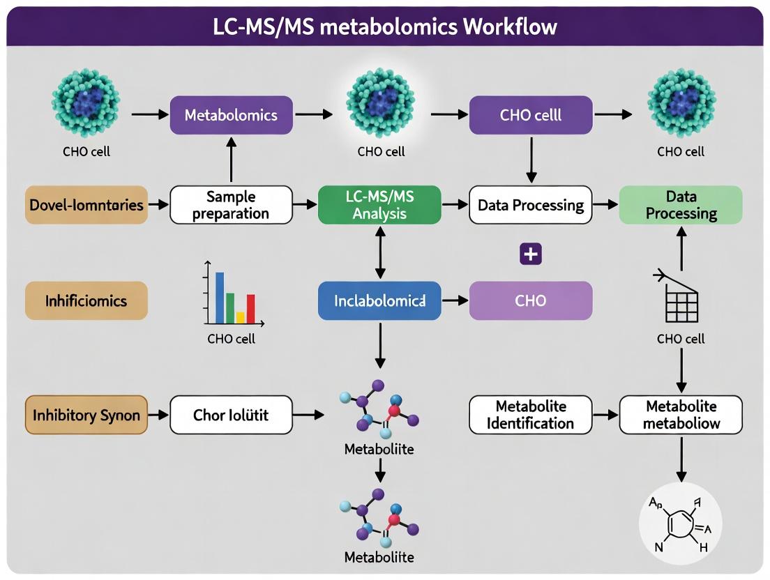
Diagrams of Metabolic Pathways and Workflow
CHO Central Metabolism & Byproduct Formation
LC-MS/MS Metabolomics Sample Workflow
The Scientist's Toolkit: Key Reagent Solutions
Table 3: Essential Materials for CHO Cell Metabolomics
| Item | Function/Benefit |
|---|---|
| Quenching Solution (Cold 100% Methanol, -80°C) | Instantly halts enzymatic activity, "freezing" the metabolic state at time of sampling. |
| LC-MS Grade Water & Solvents (MeOH, ACN) | Minimizes background chemical noise and ion suppression during MS analysis. |
| Ammonium Acetate / Ammonium Hydroxide (LC-MS Grade) | Provides volatile buffer for HILIC chromatography at basic pH for anion separation. |
| Formic Acid (LC-MS Grade, 0.1%) | Provides volatile acid for mobile phase, aids in protonation for positive ion mode ESI. |
| Polar Metabolite Standard Mix | Contains stable isotope-labeled internal standards (e.g., ¹³C-Lactate, ¹⁵N-Glutamine) for accurate quantification. |
| HILIC Chromatography Column (e.g., BEH Amide) | Retains and separates highly polar metabolites that are poorly retained on reversed-phase columns. |
| Rapid Filtration Kit (for intracellular work) | Enables sub-second quenching of suspension cultures, more accurate than direct centrifugation. |
| Vacuum Concentrator (SpeedVac) with Cold Trap | Gently removes extraction solvents without heat-induced degradation of labile metabolites. |
Application Notes
In the context of a thesis employing LC-MS/MS metabolomics to identify novel inhibitory metabolites in Chinese Hamster Ovary (CHO) cell cultures, defining and characterizing these compounds is critical for optimizing bioprocesses and recombinant protein yields. This document provides a foundational analysis of classical inhibitors (lactate, ammonia) and a framework for discovering new candidates.
Classical Inhibitory Metabolites: A Quantitative Summary Lactate and ammonia are well-documented for their dose-dependent inhibitory effects on cell growth, viability, and protein production. The table below summarizes key quantitative data from recent studies.
Table 1: Inhibitory Effects of Lactate and Ammonia in CHO Cell Cultures
| Metabolite | Typical Inhibitory Threshold (mM) | Primary Impact on CHO Cells | Proposed Mechanism(s) |
|---|---|---|---|
| Lactate | 20 - 40 mM | Reduced specific growth rate (<50%), decreased viability, altered glycosylation. | Intracellular acidification, osmotic stress, inhibition of glycolysis, induction of apoptosis. |
| Ammonia (NH₄⁺) | 2 - 5 mM | Decreased peak VCD, reduced specific productivity, increased lactate dehydrogenase (LDH) release. | Dysregulation of intracellular pH, altered UDP-sugar pools affecting glycosylation, increased endoplasmic reticulum (ER) stress. |
| Synergistic Effect | Lactate >20mM + NH₄⁺ >3mM | Severe reduction in integrated viable cell density (IVCD >60% reduction) and titer. | Amplified ER stress and apoptotic signaling pathways. |
The Quest for Novel Inhibitory Metabolites Beyond lactate and ammonia, LC-MS/MS-based metabolomic profiling of fed-batch and perfusion cultures reveals other accumulating metabolites with potential inhibitory roles. Candidates include:
- Branched-Chain Amino Acids (BCAAs): Leucine, isoleucine, and valine can accumulate to high millimolar ranges, potentially saturating transporters and inducing metabolic reprogramming.
- Methylglyoxal (MG): A reactive dicarbonyl byproduct of glycolysis linked to advanced glycation end-product (AGE) formation and cellular stress.
- Aromatic Amino Acids: Tryptophan and phenylalanine metabolites (e.g., kynurenine) can be immunomodulatory and cytotoxic at high levels.
- Unknown Features: Non-targeted LC-MS/MS often reveals masses not in standard libraries, necessitating orthogonal identification (MSⁿ, NMR).
Key Signaling Pathways Implicated in Metabolite-Induced Inhibition The inhibitory effects of classical and novel metabolites often converge on integrated stress response and apoptosis pathways.
Experimental Protocols
Protocol 1: LC-MS/MS-Based Metabolomics for Inhibitory Metabolite Profiling
Objective: To quantitatively profile polar metabolites (including lactate, ammonia, amino acids, TCA intermediates) in CHO cell culture supernatant for identification of inhibitory candidates.
Materials: See "The Scientist's Toolkit" below. Procedure:
- Sample Quenching & Extraction:
- Rapidly collect 1 mL culture broth and quench in 4 mL of -20°C 40:40:20 Methanol:Acetonitrile:Water.
- Vortex vigorously for 30 seconds.
- Incubate at -20°C for 1 hour.
- Centrifuge at 16,000 x g for 15 minutes at 4°C.
- Transfer 4 mL of supernatant to a fresh tube and dry in a vacuum concentrator.
- Reconstitute dried pellet in 100 µL LC-MS grade water for analysis.
Liquid Chromatography (HILIC):
- Column: ZIC-pHILIC (5 µm, 150 x 4.6 mm) or equivalent.
- Mobile Phase: A = 20 mM ammonium carbonate in water (pH 9.2), B = Acetonitrile.
- Gradient: 0 min: 80% B, 15 min: 20% B, 18 min: 20% B, 18.1 min: 80% B, 25 min: 80% B.
- Flow Rate: 0.3 mL/min. Temperature: 40°C. Injection Volume: 10 µL.
Tandem Mass Spectrometry (MS/MS):
- Ionization: Heated Electrospray Ionization (H-ESI), negative polarity.
- Detection: Parallel Reaction Monitoring (PRM) or Scheduled Selected Reaction Monitoring (sSRM).
- Source Parameters: Sheath Gas: 40, Aux Gas: 10, Sweep Gas: 2, Spray Voltage: -2.8 kV, Capillary Temp: 325°C.
- Acquire data for a target list of ~150 metabolites. Include internal standards for quantification.
Data Analysis:
- Process raw files using vendor (e.g., Xcalibur QuanBrowser, Skyline) or open-source software (MRMkit).
- Perform peak integration and correct using internal standards.
- Generate concentration curves from external calibration standards.
- Correlate metabolite trajectories (especially late-stage accumulators) with drops in cell growth and productivity metrics.
Protocol 2: Functional Validation of Novel Inhibitory Metabolites
Objective: To test the direct inhibitory effect of a candidate metabolite identified via LC-MS/MS profiling.
Procedure:
- Spike-In Experiment Design:
- Prepare a concentrated stock solution of the candidate metabolite in the appropriate solvent (e.g., PBS, culture medium).
- Seed CHO cells in 24-well plates at a standard density (e.g., 0.5 x 10^6 cells/mL).
- At mid-exponential phase (e.g., Day 2), spike in the candidate metabolite to achieve a range of concentrations (e.g., 1x, 2x, 5x the observed peak culture level). Include a vehicle control.
Monitoring Cellular Response:
- Monitor cell count and viability via trypan blue exclusion daily for 3-5 days post-spike.
- At 24h and 72h post-spike, collect supernatant for metabolite analysis (Protocol 1) and product titer analysis (e.g., Protein A HPLC).
- At 48h post-spike, harvest cells for apoptosis assessment via flow cytometry using Annexin V/PI staining.
Data Interpretation:
- Plot growth curves, final titer, and % apoptotic cells against metabolite concentration.
- A dose-dependent negative response confirms an inhibitory role. Compare the potency (IC50 for growth) to lactate and ammonia.
The Scientist's Toolkit: Research Reagent Solutions
Table 2: Essential Materials for Inhibitory Metabolite Research
| Item | Function & Rationale | Example Product/Catalog |
|---|---|---|
| ZIC-pHILIC LC Column | Provides robust separation of polar, ionic metabolites (lactate, amino acids, nucleotides) for MS detection. | Merck SeQuant ZIC-pHILIC (150 x 4.6 mm, 5 µm). |
| Stable Isotope-Labeled Internal Standards | Enables accurate quantification by correcting for matrix-induced ionization suppression in MS. | Cambridge Isotope Laboratories CLM-1576 (13C6-Glucose), MSK-A2-1.2 (15N-Amino Acid Mix). |
| Commercial Metabolite Calibration Standard | Contains precise mixes of target analytes for generating external calibration curves. | Biocrates MIX-STD IROA Technologies MSQLC-100. |
| Apoptosis Detection Kit | Quantifies early/late apoptosis and necrosis following metabolite exposure. | Thermo Fisher Annexin V-FITC/PI Kit (V13242). |
| CHO Cell Chemically Defined Medium | Consistent, animal-component-free baseline for reproducible spike-in experiments. | Gibco CHO CD FortiCHO or comparable. |
| Ammonium Chloride (NH₄Cl) Solution | Used as a positive control inhibitor in spike-in validation experiments. | Sigma Aldrich A9434 (1M solution in H2O). |
| Sodium L-Lactate Solution | Used as a positive control inhibitor in spike-in validation experiments. | Sigma Aldrich 71718 (≥98%, ~60% w/w syrup). |
The Impact of Metabolite Accumulation on Cell Viability, Growth, and Recombinant Protein Titer
Within the context of a broader LC-MS/MS metabolomics thesis aimed at identifying novel inhibitory metabolites in Chinese Hamster Ovary (CHO) cell cultures, this application note details the critical impact of metabolite accumulation on bioprocess performance. The accumulation of both metabolic by-products (e.g., lactate, ammonia) and novel inhibitory metabolites identified via untargeted metabolomics can severely impair cell viability, inhibit growth, and diminish recombinant protein titer. Monitoring and mitigating these effects is essential for optimizing biopharmaceutical manufacturing.
Key Quantitative Data on Inhibitory Metabolites
Table 1: Impact of Common and Novel Metabolites on CHO Cell Culture Performance
| Metabolite | Typical Accumulation Range (mM) | Reduction in Viability (%) | Reduction in Specific Growth Rate (%) | Impact on Titer (%) | Primary Identification Method |
|---|---|---|---|---|---|
| Lactate | 20 - 100 | 15-40 | 20-50 | -25 to -60 | Biochemical Analyzer / LC-MS |
| Ammonia | 2 - 10 | 10-30 | 15-40 | -20 to -50 | Ion-Selective Electrode / LC-MS |
| Methylglyoxal* | 0.005 - 0.05 | 25-60 | 30-65 | -30 to -70 | LC-MS/MS (Derivatized) |
| Choline* | 5 - 20 | 5-20 | 10-30 | -10 to -40 | LC-MS/MS |
| Unknown Metabolite X* | N/A | 20-50 | 25-55 | -35 to -65 | LC-MS/MS, NMR |
Examples of metabolites identified via advanced LC-MS/MS metabolomics screens. *Hypothetical data for a novel inhibitory metabolite; requires empirical confirmation.
Experimental Protocols
Protocol 1: LC-MS/MS-Based Metabolomics for Identifying Inhibitory Metabolites
Objective: To perform untargeted metabolomics on spent CHO cell culture media to identify novel accumulating metabolites correlating with decreased performance.
Materials:
- CHO cells in fed-batch culture.
- Quenching Solution: 60% methanol buffered with 5 mM HEPES (pH 7.4, -40°C).
- Extraction Solvent: 80% methanol/water (-80°C).
- Internal Standards: e.g., isotopically labeled amino acids, nucleotides.
- LC-MS/MS system (Q-TOF or Orbitrap preferred).
- Reversed-phase (C18) and HILIC chromatography columns.
Procedure:
- Sample Collection: Collect culture supernatant at 24-hour intervals. Immediately centrifuge (1000 x g, 5 min, 4°C) to remove cells.
- Metabolite Extraction: Mix 50 µL of supernatant with 200 µL of cold extraction solvent. Vortex vigorously for 1 min. Incubate at -80°C for 1 hour.
- Protein Precipitation: Centrifuge at 14,000 x g for 15 min at 4°C. Transfer 200 µL of supernatant to a fresh tube. Dry under a gentle stream of nitrogen or vacuum.
- Reconstitution: Reconstitute the dried extract in 100 µL of 5% acetonitrile/water for RP-LC or 100 µL acetonitrile/water (90:10) for HILIC.
- LC-MS/MS Analysis:
- RP-LC: Use a C18 column with mobile phase A (0.1% formic acid in water) and B (0.1% formic acid in acetonitrile). Apply a gradient from 5% to 95% B over 15-20 min.
- HILIC: Use an amide or silica column with mobile phase A (10 mM ammonium acetate in 90% acetonitrile, pH 9) and B (10 mM ammonium acetate in water, pH 9). Apply a gradient from high A to high B.
- Acquire data in both positive and negative electrospray ionization modes with data-dependent acquisition (DDA) or data-independent acquisition (DIA).
- Data Processing: Use software (e.g., Compound Discoverer, XCMS, MaxQuant) for peak picking, alignment, and compound identification against databases (HMDB, METLIN, in-house libraries).
Protocol 2: Functional Validation of Candidate Inhibitory Metabolites
Objective: To test the direct impact of metabolites identified in Protocol 1 on CHO cell health and productivity.
Materials:
- CHO-S or CHO-K1 suspension cells.
- Proprietary basal and feed media.
- Candidate metabolite (e.g., methylglyoxal, choline, or purified "Metabolite X").
- Bioreactor or shake flask platform.
- Trypan Blue or automated cell counter.
- Titer assay (e.g., Protein A HPLC, Octet).
Procedure:
- Spike-In Experiment Design: Set up 125 mL shake flasks with 30 mL of culture at a seeding density of 3e5 cells/mL.
- Prepare a concentration gradient of the candidate metabolite (e.g., 0x, 0.5x, 1x, 2x the peak concentration observed in production cultures).
- Culture Monitoring: Culture flasks at 36.5°C, 5% CO2, 120 rpm. Sample daily for:
- Viable Cell Density (VCD) & Viability: Using Trypan Blue exclusion.
- Metabolite Analysis: Monitor glucose, lactate, ammonia, and the candidate metabolite via biochemical analyzer or LC-MS.
- Titer Analysis: Quantify recombinant protein concentration from clarified supernatant.
- Data Analysis: Calculate specific growth rate (µ), integral viable cell count (IVCC), and specific productivity (qP). Correlative and dose-response analysis will confirm the inhibitory role of the metabolite.
Visualization of Workflow and Pathway
Title: Workflow for Identifying Inhibitory Metabolites via LC-MS/MS
Title: How Metabolite Accumulation Impacts Cell Culture Performance
The Scientist's Toolkit: Key Research Reagent Solutions
Table 2: Essential Materials for Metabolite Impact Studies in CHO Cells
| Item / Reagent Solution | Function & Application in Research | Example Vendor(s) |
|---|---|---|
| Quenching & Extraction Kits | Standardized, cold solvent mixtures for immediate metabolic arrest and extraction, ensuring reproducibility in LC-MS sample prep. | Bioteke, Biovision, MilliporeSigma |
| LC-MS/MS Grade Solvents | Ultra-pure solvents (water, methanol, acetonitrile, formic acid) to minimize background noise and ion suppression during metabolomics analysis. | Fisher Chemical, Honeywell |
| Stable Isotope-Labeled Internal Standards | Isotopically labeled amino acids, organic acids, and nucleotides for absolute quantification and monitoring extraction efficiency in complex media. | Cambridge Isotope Labs, Sigma-Isotec |
| CHO Chemically Defined Media | Animal-component-free, consistent basal and feed media essential for controlled spike-in validation studies without confounding variables. | Gibco (Thermo Fisher), Irvine Scientific |
| Automated Cell Counters & Viability Analyzers | Instruments for rapid, accurate daily monitoring of Viable Cell Density (VCD) and viability (via Trypan Blue). | Bio-Rad (TC20), Nexcelom (Cellometer), Invitrogen (Countess) |
| Microplate-Based Metabolite Assays | Colorimetric/Fluorometric kits for rapid screening of key metabolites (glucose, lactate, glutamate, ammonia) in culture supernatant. | BioVision, Abcam, Promega |
| Recombinant Protein Titer Assay Kits | Pre-optimized kits (e.g., Protein A HPLC, SimpleStep ELISA, Octet Dip-and-Read) for high-throughput, accurate titer measurement. | Protein A Biosensors (Sartorius), Cisbio, ProteinSimple |
| High-Resolution Mass Spectrometer | Q-TOF or Orbitrap systems coupled to UHPLC for high-sensitivity, high-resolution untargeted metabolomics and identification of novel metabolites. | Sciex (TripleTOF), Thermo (Q Exactive), Agilent (6546 LC/Q-TOF) |
The Role of LC-MS/MS Metabolomics in Unbiased Metabolic Phenotyping
Within the thesis investigating LC-MS/MS metabolomics for identifying novel inhibitory metabolites in Chinese Hamster Ovary (CHO) cell cultures, unbiased metabolic phenotyping stands as the critical discovery engine. This approach provides a global, non-targeted view of the metabolome, enabling the identification of previously uncharacterized metabolites that accumulate under specific process conditions and potentially inhibit cell growth or recombinant protein production. LC-MS/MS combines the superior separation power of liquid chromatography with the high sensitivity and structural elucidation capabilities of tandem mass spectrometry, making it the platform of choice for this endeavor.
Key Applications in CHO Cell Research
| Application Area | Primary Objective | Typical LC-MS/MS Configuration | Key Metabolite Classes Identified |
|---|---|---|---|
| Process Optimization | Identify metabolites correlating with high/low titer or cell viability. | HILIC & Reversed-Phase, Q-TOF or Orbitrap | Organic acids, nucleotides, amino acids, lipids |
| Media Analysis | Uncover inhibitory components in spent media. | Reversed-Phase (C18), Triple Quadrupole for profiling | Lactate, ammonium, Ala-Arg, other dipeptides |
| Cell Engineering Assessment | Phenotype of engineered cell lines (e.g., apoptosis-resistant). | Dual LC separation, High-res MS/MS | TCA cycle intermediates, redox cofactors, caspase substrates |
| Clone Selection | Find metabolic signatures of high-producing clones early in bioprocessing. | Rapid LC gradients, High-throughput MS | Energy charge metabolites, glycosylation precursors |
| Quantitative Data from Representative CHO Cell Metabolomics Studies | |||
|---|---|---|---|
| Study Focus: Lactate Shift | |||
| Metabolite: Lactate | Concentration (High-Lactate Phenotype): 25-35 mM | Concentration (Low-Lactate Phenotype): <5 mM | Impact: High lactate correlates with osmolality stress and inhibited growth. |
| Study Focus: Unknown Inhibitor | |||
| Metabolite: Tryptophan-N(oxide) | Fold-Change in Spent Media: 15x | Identified by: Accurate mass (<5 ppm) & MS/MS library match | Postulated Effect: Linked to oxidative stress response. |
| Study Focus: Energy Metabolism | |||
| Metabolite: ATP/ADP Ratio | Value in Proliferating Phase: 8-12 | Value in Stationary/Decline Phase: 2-4 | Significance: Indicator of metabolic stress and energetic state. |
Detailed Protocols
Protocol 1: Sample Preparation for Intracellular Metabolites from CHO Cells
Goal: Quench metabolism and extract polar and semi-polar metabolites for unbiased LC-MS/MS analysis.
Materials:
- CHO cell culture (e.g., in bioreactor or shake flask)
- Quenching Solution: 60% methanol (HPLC grade) in water, pre-chilled to -40°C
- Extraction Solvent: 80% methanol/water with internal standards (e.g., ( ^{13}C ), ( ^{15}N )-labeled amino acids), -20°C
- PBS (phosphate-buffered saline), 4°C
- Cell scraper (for adherent cells) or centrifuge (for suspension)
Procedure:
- Quenching: Rapidly transfer 1 mL of cell culture (~1-2x10^7 cells) into 4 mL of cold quenching solution. Vortex immediately. Incubate on dry ice or at -40°C for 10 min.
- Washing: Centrifuge at 4000 x g at -10°C for 10 min. Discard supernatant.
- Extraction: Resuspend cell pellet in 1 mL of cold extraction solvent. Vortex vigorously for 30 sec.
- Processing: Sonicate on ice for 5 min, then shake at 4°C for 30 min.
- Clearing: Centrifuge at 16,000 x g at 4°C for 15 min.
- Storage: Transfer supernatant (metabolite extract) to a fresh tube. Dry under a gentle stream of nitrogen or in a vacuum concentrator. Store dried extract at -80°C until LC-MS/MS analysis.
- Reconstitution: Prior to analysis, reconstitute in 100 µL of appropriate LC starting solvent (e.g., 2% acetonitrile/water for HILIC, or water for RP).
Protocol 2: Untargeted LC-MS/MS Analysis for Metabolic Phenotyping
Goal: Acquire comprehensive MS1 and data-dependent MS2 (dd-MS2) spectra for metabolite identification.
LC Conditions (HILIC for Polar Metabolites):
- Column: SeQuant ZIC-pHILIC (5 µm, 150 x 4.6 mm)
- Mobile Phase A: 20 mM ammonium carbonate in water, pH 9.2
- Mobile Phase B: Acetonitrile
- Gradient: 80% B to 20% B over 20 min, hold 5 min, re-equilibrate.
- Flow Rate: 0.3 mL/min
- Column Temp: 40°C
- Injection Volume: 10 µL
MS Conditions (High-Resolution Q-TOF or Orbitrap):
- Ionization: ESI, positive and negative polarity modes (separate runs)
- Mass Range (MS1): 70-1200 m/z
- Resolution: >30,000 FWHM
- dd-MS2 Settings: Top 10 most intense ions per cycle, exclude after 2 times for 30 sec.
- Collision Energy: Stepped (e.g., 20, 40, 60 eV)
Data Processing:
- Use software (e.g., MS-DIAL, Compound Discoverer, XCMS) for peak picking, alignment, and deconvolution.
- Annotate features using accurate mass (±5 ppm) and MS/MS fragmentation against databases (HMDB, METLIN, NIST, or in-house CHO-specific library).
- Perform statistical analysis (PCA, PLS-DA) to identify significant features (VIP >1.5, p-value <0.05) distinguishing sample groups.
Protocol 3: Identification and Validation of Novel Inhibitory Metabolites
Goal: Confirm the structure and test the inhibitory effect of a candidate metabolite identified from untargeted screening.
Materials:
- Purified candidate metabolite (commercial or synthesized)
- Basal cell culture media
- Viability assay kit (e.g., Trypan blue, MTT)
Procedure:
- Spike-In Experiment: Prepare fresh culture media spiked with a concentration gradient of the candidate metabolite (e.g., 0 µM, 10 µM, 50 µM, 200 µM).
- Cell Treatment: Seed CHO cells at standard density in media containing the metabolite. Include a vehicle control.
- Monitoring: Monitor cell growth, viability, and specific productivity over 3-5 days.
- Metabolite Profiling: At a key time point, harvest cells and media from treated and control cultures. Perform targeted LC-MS/MS (MRM mode on a triple quadrupole) to quantify the candidate and related pathway metabolites.
- Data Integration: Correlate the intracellular/extracellular concentration of the candidate with the observed phenotypic inhibition. Use pathway analysis tools (e.g., Mummichog, MetaboAnalyst) to map the metabolite onto metabolic pathways and predict its origin and impact.
The Scientist's Toolkit: Research Reagent Solutions
| Item | Function in CHO Metabolomics |
|---|---|
| Quenching Solution (Cold 60% MeOH) | Instantly halts enzymatic activity to "snapshot" the intracellular metabolic state. |
| Stable Isotope-Labeled Internal Standards (e.g., ( ^{13}C_6 )-Glucose) | Enables absolute quantification and correction for ionization efficiency variation during MS analysis. |
| HILIC Chromatography Column | Effectively retains and separates highly polar metabolites (e.g., nucleotides, CoAs) poorly retained by reversed-phase. |
| High-Resolution Mass Spectrometer (Orbitrap/Q-TOF) | Provides accurate mass measurement for elemental composition determination and confident database matching. |
| Metabolomics Databases (HMDB, METLIN) | Public spectral libraries for matching MS/MS fragmentation patterns to putative metabolite identities. |
| Cell Culture Bioreactor with Online Sensors | Allows for correlated analysis of metabolic profiles with process parameters (pH, pO2, viability). |
| Pathway Analysis Software (MetaboAnalyst) | Statistically enriches significant metabolite changes into biological pathways for functional interpretation. |
Workflow for Unbiased Phenotyping in CHO Research
Pathway Context for Novel Inhibitor Discovery
From Cell Culture to Candidate List: A Step-by-Step LC-MS/MS Metabolomics Workflow for Inhibitor Discovery
Within the broader thesis on applying LC-MS/MS metabolomics to identify novel inhibitory metabolites in Chinese Hamster Ovary (CHO) cell bioprocessing, this experimental design is fundamental. The core hypothesis is that metabolic bottlenecks, manifesting as accumulations of inhibitory metabolites, differentially characterize low-performing cultures or specific fed-batch time points. A direct comparative analysis between high- and low-performing conditions enables the pinpointing of these critical metabolic differences, moving beyond correlative observations to identify causative factors in cell culture performance decline.
Core Experimental Design Frameworks
Two primary, complementary frameworks are employed:
A. Performance-Based Comparison (Endpoint Analysis):
- Design: Cultivate multiple bioreactors under standardized conditions. At harvest, classify cultures into "High" (e.g., >5 g/L final titer, >90% viability at day 14) and "Low" (e.g., <3 g/L, <70% viability) performance groups.
- Sampling: Collect cells and spent media at matched time points (e.g., exponential growth phase, late production phase) from each bioreactor for integrated metabolomics.
- Objective: Identify metabolic signatures consistently associated with the high- or low-performance state, irrespective of chronological time.
B. Temporal Trajectory Comparison (Fed-Batch Time Series):
- Design: Monitor a single or multiple replicate fed-batch runs intensively over time.
- Sampling: Collect frequent samples (e.g., every 12-24 hours) from the same bioreactor(s) throughout the run.
- Comparison Logic: Analyze time points representing early high-performance (e.g., day 3-5, peak growth) versus late declining-performance (e.g., day 12-14, onset of rapid apoptosis).
- Objective: Uncover the dynamic metabolic shifts that precede and accompany performance decline, revealing the metabolic trajectory into inhibition.
Detailed Protocols for LC-MS/MS Metabolomics Workflow
Protocol 3.1: Quenching, Extraction, and Sample Preparation for Intracellular Metabolites
Principle: Rapidly halt metabolism and extract polar and semi-polar metabolites.
- Quenching: Withdraw culture broth (e.g., 5 mL) and immediately syringe it into 20 mL of pre-chilled (-40°C) 60% methanol/water quenching solution. Vortex.
- Centrifugation: Pellet cells at 4000 x g for 5 min at -20°C. Discard supernatant.
- Wash: Resuspend cell pellet in 5 mL cold PBS. Re-centrifuge. Decant supernatant.
- Extraction: Add 1 mL of pre-chilled (-40°C) 80% methanol/water (v/v) containing internal standards (e.g., ( ^{13}\text{C} )-labeled amino acids, ( ^{15}\text{N} )-labeled nucleotides) to the pellet. Vortex vigorously for 30s.
- Incubation: Place on dry ice for 15 min, then in a -20°C freezer for 1 hour.
- Clarification: Centrifuge at 16,000 x g for 15 min at 4°C. Transfer supernatant (metabolite extract) to a fresh tube.
- Drying: Dry extracts in a vacuum concentrator without heat.
- Reconstitution: Reconstitute in 100 µL of LC-MS grade water for hydrophilic interaction liquid chromatography (HILIC) or 5% methanol for reversed-phase analysis. Vortex, centrifuge, and transfer to LC-MS vials.
Protocol 3.2: Extracellular Metabolite (Spent Media) Preparation
Principle: Deplete proteins and stabilize metabolites in spent media.
- Clarification: Centrifuge 1 mL of culture broth at 16,000 x g for 10 min at 4°C to remove cells and debris.
- Protein Precipitation: Transfer 500 µL of supernatant to a tube containing 1500 µL of cold (-20°C) 100% methanol (containing internal standards). Vortex for 1 min.
- Incubation: Hold at -20°C for 1 hour.
- Centrifugation: Centrifuge at 16,000 x g for 15 min at 4°C.
- Transfer & Dry: Transfer supernatant to a new tube. Dry under vacuum.
- Reconstitution: Reconstitute in 100 µL of LC-MS starting mobile phase (e.g., 95:5 H₂O:ACN for HILIC). Centrifuge and analyze.
Protocol 3.3: LC-MS/MS Analysis Parameters (HILIC-Positive Mode)
- LC System: UHPLC with autosampler maintained at 4°C.
- Column: ZIC-pHILIC (150 x 2.1 mm, 5 µm) or equivalent.
- Mobile Phase: A = 20 mM ammonium carbonate, 0.1% ammonium hydroxide (pH ~9.2); B = Acetonitrile.
- Gradient:
- 0-2 min: 80% B
- 2-17 min: 80% B → 20% B
- 17-18 min: 20% B
- 18-18.5 min: 20% B → 80% B
- 18.5-25 min: 80% B (re-equilibration)
- Flow Rate: 0.2 mL/min. Column Temp: 40°C.
- MS System: High-resolution tandem MS (e.g., Q-TOF, Orbitrap).
- Ionization: ESI-Positive.
- Scan Range: m/z 70-1050.
- Data Acquisition: Data-Dependent Acquisition (DDA) or Parallel Reaction Monitoring (PRM) for targeted quantification.
Table 1: Representative Metabolites Differentially Abundant in Low-Performing CHO Cultures / Late Time Points
| Metabolite Class | Metabolite Name | Observed Trend (Low vs. High / Late vs. Early) | Putative Role/Inhibitory Mechanism | p-value (example) | Fold Change |
|---|---|---|---|---|---|
| Amino Acids | Lactate | Significantly Increased | Glycolytic end-product; inhibits growth at high [ ] | <0.001 | >3.0 |
| Ammonia | Significantly Increased | Disrupts intracellular pH, UDP-GlcNAc biosynthesis | <0.001 | >2.5 | |
| TCA Cycle | Citrate | Often Decreased | Indicative of mitochondrial stress or export for lipids | 0.005 | 0.4 |
| Nucleotides | Xanthine, Hypoxanthine | Increased | Purine degradation products; potential indicators of energy stress | <0.01 | >2.0 |
| Lipid-Related | Choline | Accumulates | Possible phospholipid turnover or transport alteration | 0.02 | 1.8 |
| Acylcarnitines (C14, C16) | Increased | Suggests incomplete fatty acid oxidation | <0.01 | >2.2 | |
| Novel Candidates | N-Acetylputrescine | Markedly Increased | Polyamine derivative; may correlate with apoptosis | <0.001 | >5.0 |
| UDP-sugars (e.g., UDP-Glc) | Decreased | Linked to ER stress and altered glycosylation | 0.01 | 0.5 |
Visualization of Pathways and Workflows
Title: LC-MS Metabolomics Workflow for CHO Performance Comparison
Title: Inferred Metabolic Dysregulation in Low-Performance CHO Cultures
The Scientist's Toolkit: Research Reagent Solutions
Table 2: Essential Materials for Comparative CHO Cell Metabolomics
| Item / Reagent Solution | Function & Importance |
|---|---|
| Quenching Solution (60% Methanol, -40°C) | Instantly halts enzymatic activity, "freezing" the metabolic state at sampling time. Critical for accurate intracellular snapshots. |
| Extraction Solvent (80% Methanol with ISTDs) | Efficiently lyses cells and extracts a broad range of polar metabolites. Inclusion of isotope-labeled internal standards corrects for ion suppression and variability. |
| Stable Isotope-Labeled Internal Standard Mix | A cocktail of ( ^{13}\text{C} ), ( ^{15}\text{N} )-labeled amino acids, nucleotides, and central carbon metabolites. Essential for semi-quantitative comparison and data normalization. |
| HILIC Chromatography Column | Enables retention and separation of highly polar metabolites (sugars, organic acids, amino acids) that elute early or not at all on reversed-phase columns. |
| Ammonium Carbonate/Ammonia Buffers | Volatile buffers for HILIC-MS that provide optimal pH for separation and are MS-compatible (leave no residue in source). |
| Quality Control (QC) Pool Sample | A composite of all experimental samples, injected repeatedly throughout the run. Monitors instrument stability and is used for data correction (e.g., LOESS signal correction). |
| Metabolomics Software Suite | Tools like Compound Discoverer, XCMS, or Skyline for peak picking, alignment, and statistical analysis. Required for handling large, complex datasets. |
| Metabolite Database (e.g., HMDB, mzCloud) | Spectral libraries for annotating detected m/z features. Critical for moving from unknown peaks to identified metabolites and pathways. |
This protocol details robust methodologies for the preparation of intracellular metabolite samples from Chinese Hamster Ovary (CHO) cells, a critical pre-analytical phase for LC-MS/MS metabolomics. Within the broader thesis aim of identifying novel inhibitory metabolites in CHO cell bioprocessing, consistent and accurate sample preparation is paramount. The goal is to rapidly arrest metabolism (quenching), efficiently extract a broad spectrum of metabolites, and apply normalization strategies to enable biologically meaningful comparative analysis via LC-MS/MS.
Core Principles and Critical Considerations
- Speed is Critical: Metabolic turnover can occur in seconds. The interval between quenching and full extraction must be minimized.
- Temperature Control: Maintain cold temperatures (using dry ice, liquid nitrogen, or ice-cold solvents) throughout quenching and extraction to inhibit enzymatic activity.
- Completeness vs. Selectivity: Extraction solvents must balance the breadth of metabolite classes (polar, semi-polar, ionic) with the selectivity required for subsequent LC-MS/MS analysis.
- Minimal Sample Handling: Reduce steps to minimize metabolite loss, adsorption, or degradation.
- Integration with Broader Thesis: These protocols are designed to capture metabolite snapshots that can be correlated with cell culture performance data (e.g., viability, titer, nutrient consumption) to pinpoint metabolites associated with growth inhibition or productivity bottlenecks.
Detailed Protocols
Protocol 3.1: Rapid Filtration & Cold Methanol Quenching
This method is preferred for adherent or suspension CHO cells to rapidly separate cells from nutrient-rich media, which can interfere with intracellular measurements.
Materials:
- CHO cell culture
- Vacuum filtration manifold
- Cold phosphate-buffered saline (PBS, 4°C)
- Quenching Solution: 60% aqueous methanol (HPLC grade) kept at -40°C to -50°C (dry ice/ethanol bath).
- Cell scraper (for adherent cells)
- Liquid nitrogen
Procedure:
- Preparation: Pre-chill filtration manifold and quenching solution. Label collection tubes.
- Harvest: For suspension cells, rapidly transfer culture aliquot onto a pre-wetted, cold membrane filter under gentle vacuum (<15 kPa). For adherent cells, quickly aspirate media, rinse with cold PBS, scrape into cold PBS, and filter.
- Wash: Immediately wash cells on filter with 10 mL of ice-cold PBS.
- Quench: Within 10 seconds of washing, apply 5 mL of cold (-40°C) 60% methanol quenching solution to the cell bed. Simultaneously, use a pre-chilled spatula to scrape the cell paste into a 2 mL tube containing 1 mL of quenching solution, submerged in liquid nitrogen. Flash-freeze.
- Storage: Store samples at -80°C until extraction.
Protocol 3.2: Metabolite Extraction via Cold Solvent Partition
This biphasic extraction method efficiently recovers a wide range of metabolites.
Materials:
- Quenched cell pellets (from Protocol 3.1)
- Extraction Solvent I: Cold (-20°C) Methanol (100%)
- Extraction Solvent II: Cold (-20°C) Water
- Extraction Solvent III: Cold (-20°C) Chloroform
- Sonicator with microtip
- Centrifuge capable of 15,000 g at 4°C
- Vacuum concentrator (SpeedVac)
Procedure:
- Homogenization: To the frozen quenched pellet, add 400 µL of cold methanol and 200 µL of cold water. Vortex vigorously for 30 seconds.
- Sonication: Sonicate the mixture on ice for 2 minutes (5 sec pulse on, 10 sec off, 30% amplitude).
- Phase Separation: Add 400 µL of cold chloroform. Vortex for 1 minute.
- Incubation: Incubate the mixture at -20°C for 20 minutes.
- Centrifugation: Centrifuge at 15,000 g for 15 minutes at 4°C. This will separate the mixture into a lower organic (chloroform) phase, an interface (protein/DNA disc), and an upper aqueous phase containing polar intracellular metabolites.
- Collection: Carefully transfer 350 µL of the upper aqueous phase to a fresh, pre-chilled microcentrifuge tube. Avoid disturbing the interface.
- Drying: Dry the aqueous extract using a vacuum concentrator (SpeedVac) without heat (~2 hours).
- Reconstitution: Reconstitute the dried metabolite pellet in 100 µL of LC-MS compatible solvent (e.g., 5% acetonitrile in water) matched to the starting conditions of your chromatographic method. Vortex for 30 seconds and centrifuge at 15,000 g for 10 minutes at 4°C.
- Final Storage: Transfer the supernatant to an LC-MS vial. Store at -80°C until analysis.
Protocol 3.3: Sample Normalization Strategies
Accurate normalization is required to correct for variations in cell number or biomass prior to comparative analysis.
Strategy A: Pre-Quenching Cell Count Normalization
- Count cells from a parallel sample using an automated cell counter or hemocytometer.
- Harvest a volume of culture containing exactly 2 x 10^6 cells for quenching and extraction.
- Perform extraction (Protocol 3.2) and reconstitute in a fixed volume (e.g., 100 µL). Data are directly comparable per 10^6 cells.
Strategy B: Post-Extraction Biomass Proxy Normalization
- During the extraction (Protocol 3.2, Step 5), after collecting the aqueous phase, allow the protein disc at the interface to air dry.
- Redissolve the protein pellet in 200 µL of 0.1M NaOH by heating at 95°C for 10 minutes.
- Quantify the total protein content using a microplate Bradford or BCA assay.
- Normalize the metabolite LC-MS peak areas to the total protein amount (e.g., per µg of protein).
Strategy C: Internal Standard (IS) Normalization
- Add a known amount of a non-naturally occurring internal standard (e.g., deuterated or 13C-labeled metabolites like d27-myristic acid or 13C6-glucose) during the reconstitution step (Protocol 3.2, Step 8).
- Use the stable signal of the IS across all samples to correct for instrument variability. Note: This corrects for analytical drift, not biological differences in biomass.
Data Tables
Table 1: Comparison of Quenching Methods for CHO Cells
| Quenching Method | Principle | Advantages | Disadvantages | Suitability for Thesis |
|---|---|---|---|---|
| Cold Methanol (-40°C) | Rapid thermal/enzymatic arrest | Fast, effective for many pathways, compatible with filtration. | Can cause cell leakage for some cell types. | High. Robust and widely validated for suspension CHO. |
| Liquid N2 Freezing | Instant freezing | Gold standard for arresting metabolism. | Requires rapid handling, not always feasible for large sample sets. | Medium. Best for small-scale, high-value samples. |
| Acid Treatment (e.g., PCA) | pH denaturation of enzymes | Very effective quenching. | Requires neutralization, introduces salts. | Low. Adds complexity, risk of metabolite degradation. |
Table 2: Common Extraction Solvent Systems for Intracellular Metabolomics
| Solvent System | Phase | Target Metabolite Classes | Key Consideration |
|---|---|---|---|
| Methanol/Water/Chloroform | Biphasic | Aqueous: Amino acids, sugars, organic acids, nucleotides. Organic: Lipids, acyl-CoAs. | Comprehensive coverage; separates polar/non-polar. |
| Acetonitrile/Methanol/Water | Monophasic | Broad polar and semi-polar metabolites. | Simpler protocol; good for hydrophilic interaction LC. |
| Methanol/Water | Monophasic | Highly polar, central carbon metabolites. | May miss less polar metabolites. |
Table 3: Normalization Methods for Comparative Analysis
| Normalization Method | What it Corrects For | Procedure Point | Data Output |
|---|---|---|---|
| Cell Counting | Differences in cell number per sample. | Pre-harvest. | Metabolite abundance per 10^6 cells. |
| Total Protein | Differences in total biomass. | Post-extraction. | Metabolite abundance per µg protein. |
| DNA Quantification | Differences in cell number. | Post-extraction. | Metabolite abundance per µg DNA. |
| Internal Standard (IS) | Instrumental variance, injection error. | Reconstitution/Analysis. | Peak Area Ratio (Analyte/IS). |
| Sample Median | Global systemic shifts. | Post-acquisition (data processing). | Scaled to median sample intensity. |
Visualization: Workflow and Pathway Diagrams
Diagram Title: Comprehensive Metabolite Sample Prep Workflow
Diagram Title: Protocol Role in Broader Metabolomics Thesis
The Scientist's Toolkit: Research Reagent Solutions
| Item | Function in Protocol | Key Consideration |
|---|---|---|
| 60% Aqueous Methanol (-40°C) | Quenching solvent. Rapidly cools cells and inhibits enzyme activity. | Must be pre-chilled in dry ice/ethanol bath. Use HPLC grade solvents. |
| Chloroform (HPLC Grade) | Organic phase in extraction. Solubilizes lipids and facilitates phase separation. | Toxic; use in fume hood. Must be cold to improve metabolite recovery. |
| Deuterated Internal Standards (e.g., d27-Myristate) | Added during reconstitution. Corrects for instrumental variance and sample loss. | Should not be naturally present in samples. Spike at consistent concentration. |
| BCA or Bradford Protein Assay Kit | Quantifies protein from the extraction interface for biomass normalization. | Compatible with residual solvents; may require dilution. |
| 0.1 μm Nylon Membrane Filters | For rapid filtration of cells from media during quenching. | Low protein binding ensures minimal metabolite adhesion. |
| Cryogenic Vials & Pre-chilled Racks | For handling and flash-freezing samples in liquid nitrogen. | Maintains cold chain, prevents thawing. |
| Vacuum Concentrator (SpeedVac) | Gently removes extraction solvents without heat to prevent degradation. | Essential for reproducible metabolite drying and reconstitution. |
Within the broader thesis research focused on discovering novel inhibitory metabolites in Chinese Hamster Ovary (CHO) cell cultures for bioprocess optimization, developing a robust LC-MS/MS method is paramount. This application note details the comprehensive method development strategy aimed at achieving broad metabolite coverage. The goal is to separate and detect a wide range of endogenous metabolites—from polar amino acids and sugars to non-polar lipids—to identify those negatively impacting cell growth and recombinant protein productivity.
Core Principles for Broad Coverage
Broad-coverage metabolomics requires orthogonal approaches in both chromatography and mass spectrometry. Key principles include:
- Chromatography: Employing complementary separation mechanisms (e.g., reversed-phase and hydrophilic interaction liquid chromatography) to capture metabolites of diverse polarities.
- Mass Spectrometry: Optimizing electrospray ionization (ESI) parameters for both positive and negative modes and employing data-dependent and independent acquisition to maximize compound detection and identification.
Chromatography Method Development
Dual-Column Strategy
A single chromatographic method is insufficient. A dual-platform approach is implemented.
Platform A: Reversed-Phase Liquid Chromatography (RPLC)
- Application: Medium to non-polar metabolites (e.g., lipids, acyl-carnitines, bile acids, steroids).
- Column: C18 column with polar embedded groups (e.g., Acquity UPLC HSS T3, 2.1 x 100 mm, 1.8 µm) for better retention of moderately polar compounds.
- Mobile Phase: A: Water with 0.1% Formic Acid; B: Acetonitrile with 0.1% Formic Acid.
- Gradient: Shallow gradient for enhanced separation.
- 0-2 min: 1% B
- 2-15 min: 1-99% B
- 15-17 min: 99% B
- 17-17.1 min: 99-1% B
- 17.1-20 min: 1% B (Re-equilibration)
- Temperature: 40°C
- Flow Rate: 0.4 mL/min
Platform B: Hydrophilic Interaction Liquid Chromatography (HILIC)
- Application: Polar metabolites (e.g., amino acids, organic acids, nucleotides, sugars, phosphorylated intermediates).
- Column: Zwitterionic HILIC column (e.g., Merck SeQuant ZIC-pHILIC, 2.1 x 150 mm, 5 µm).
- Mobile Phase: A: 20 mM Ammonium Carbonate in Water, pH 9.2; B: Acetonitrile.
- Gradient:
- 0-2 min: 80% B
- 2-17 min: 80-20% B
- 17-19 min: 20% B
- 19-19.1 min: 20-80% B
- 19.1-25 min: 80% B (Re-equilibration)
- Temperature: 40°C
- Flow Rate: 0.15 mL/min
Method Comparison Table
Table 1: Comparison of Chromatographic Platforms for CHO Cell Metabolomics.
| Parameter | RPLC (Platform A) | HILIC (Platform B) |
|---|---|---|
| Target Metabolites | Lipids, Co-factors, Steroids | Central Carbon Metabolites, Amino Acids |
| Retention Mechanism | Hydrophobicity | Polarity / Hydrophilicity |
| Typical Starting Eluent | Aqueous (Polar) | Organic (Non-polar) |
| Ionization Efficiency | Often enhanced with additives | Can be suppressed by buffers |
| MS Compatibility | Excellent with volatile acids | Requires volatile buffers (e.g., NH₄Ac) |
| Gradient Direction | Low to High Organic | High to Low Organic |
Mass Spectrometry Parameter Optimization
Instrumentation & Ion Source
- System: Triple quadrupole or Q-TOF mass spectrometer with ESI source.
- Ionization Mode: Fast polarity switching between ESI+ and ESI- within a single run is critical for coverage.
- Source Parameters (Optimized on a standard mix):
- Capillary Voltage: ±3.0 kV (positive/negative)
- Source Temperature: 150°C
- Desolvation Temperature: 500°C
- Desolvation Gas Flow: 1000 L/hr (N₂)
- Cone Gas Flow: 150 L/hr
- Nebulizer Gas Pressure: 7 bar
Acquisition Methods
For Q-TOF Systems (Untargeted Profiling):
- Mode: Data-Independent Acquisition (MSE or All-Ions MS/MS).
- Scan Range: m/z 50-1200.
- Low Energy (MS) Function: Collision Energy: 6 eV.
- High Energy (MS/MS) Function: Ramped Collision Energy: 20-40 eV.
- Scan Time: 0.1 sec per function.
For Triple Quadrupole Systems (Targeted Quantification/Validation):
- Mode: Multiple Reaction Monitoring (MRM).
- Dwell Time: 5-20 ms per transition.
- Collision Energy: Optimized for each metabolite using pure standards or via predictive software.
Table 2: Optimized MS Parameters for Broad Metabolite Screening.
| Parameter | Setting (Positive Mode) | Setting (Negative Mode) | Purpose |
|---|---|---|---|
| Capillary (kV) | +3.0 | -3.0 | Ion formation |
| Cone (V) | 30 | 30 | Ion guidance into analyzer |
| Source Temp (°C) | 150 | 150 | Solvent desolvation |
| Desolvation Temp (°C) | 500 | 500 | Complete desolvation |
| Desolvation Gas (L/hr) | 1000 | 1000 | Aid desolvation |
| Acquisition Mode | DIA / MRM | DIA / MRM | Balance of coverage & sensitivity |
| Collision Energy | Ramped (10-40 eV) | Ramped (10-40 eV) | Compound fragmentation |
Detailed Experimental Protocols
Protocol 1: Sample Preparation from CHO Cell Culture
Objective: To quench metabolism and extract a comprehensive metabolite pool from adherent or suspension CHO cells. Reagents: -80°C Methanol (LC-MS grade), PBS (4°C), Water (LC-MS grade), Internal Standard Mix (e.g., labeled amino acids, nucleotides). Procedure:
- Quenching: For suspension cells, rapidly transfer 1 mL of culture to 4 mL of -80°C 60% methanol. Vortex and hold at -80°C for 15 min.
- Harvesting: Centrifuge at 14,000 g for 15 min at -9°C. Discard supernatant.
- Extraction: Resuspend cell pellet in 1 mL of -20°C extraction solvent (40:40:20 Methanol:Acetonitrile:Water with 0.1% Formic Acid). Vortex vigorously for 30 sec.
- Processing: Sonicate on ice for 5 min, then shake at 4°C for 30 min.
- Clearing: Centrifuge at 14,000 g for 15 min at 4°C.
- Preparation: Transfer supernatant to a new tube. Dry under a gentle stream of nitrogen or in a vacuum concentrator.
- Reconstitution: Reconstitute dried extract in 100 µL of starting mobile phase appropriate for the LC method (e.g., 1% ACN for RPLC or 80% ACN for HILIC). Vortex and centrifuge.
- Analysis: Transfer to an LC-MS vial with insert. Inject 5-10 µL.
Protocol 2: System Suitability and QC
Objective: To ensure method robustness and monitor instrument performance. Procedure:
- Prepare a QC Pool Sample by combining equal volumes of all study samples.
- Prepare a System Suitability Mix containing known metabolites spanning the polarity range (e.g., leucine, glutamate, succinate, AMP, caffeine, reserpine).
- Inject the suitability mix at the beginning of the sequence to check retention time stability, peak shape, and sensitivity.
- Inject the QC pool sample repeatedly (every 4-6 injections) throughout the analytical sequence.
- Monitor QC metrics: total ion chromatogram (TIC) overlay, baseline noise, and intensity of key ions. Use principal component analysis (PCA) of QC data to ensure instrumental drift is minimal.
Visualization of Workflows and Pathways
Diagram 1: CHO Cell Metabolomics LC-MS Workflow
Diagram 2: LC Method Selection Based on Polarity
The Scientist's Toolkit: Research Reagent Solutions
Table 3: Essential Materials for LC-MS/MS Metabolomics of CHO Cells.
| Item | Function & Rationale |
|---|---|
| LC-MS Grade Solvents (Water, Methanol, Acetonitrile) | Minimize background ions and noise for high-sensitivity detection. |
| Ammonium Formate/Carbonate (LC-MS Grade) | Volatile buffer salts for mobile phases; aid ionization and control pH in HILIC. |
| Formic Acid (LC-MS Grade, 0.1%) | Common mobile phase additive for RPLC; promotes protonation in ESI+. |
| Stable Isotope-Labeled Internal Standards (e.g., 13C-AA Mix) | Correct for matrix effects and extraction variability; essential for quantification. |
| Hybrid Metabolite Standards Mix | Used for system suitability, tuning, and retention time calibration across batches. |
| Solid Phase Extraction (SPE) Plates (C18 & Polymer) | For sample clean-up to remove salts & proteins, improving column lifetime. |
| Polar-Embedded C18 Column (e.g., HSS T3) | Retains a wider range of polar metabolites than standard C18. |
| Zwitterionic HILIC Column (e.g., ZIC-pHILIC) | Separates highly polar, ionic metabolites incompatible with RPLC. |
| Quality Control (QC) Pool Sample | Monitors instrumental performance and data reproducibility throughout the run. |
Application Notes and Protocols for LC-MS/MS Metabolomics in CHO Cell Research
1. Thesis Context This protocol details the critical data processing workflow for an LC-MS/MS-based metabolomics thesis focused on identifying novel inhibitory metabolites in Chinese Hamster Ovary (CHO) cells. The objective is to compare metabolomic profiles under controlled vs. stressed (e.g., nutrient depletion, inhibitory compound exposure) conditions to detect and identify differential metabolites that may play a role in cell growth inhibition or productivity bottlenecks in bioprocessing.
2. Core Data Processing Pipeline Protocol
2.1. Experimental Workflow Diagram
Diagram Title: LC-MS Metabolomics Data Processing Workflow for CHO Cells
2.2. Detailed Methodologies
Protocol 2.2.1: Peak Picking and Feature Detection
- Software: Use MS-DIAL, MZmine 3, or XCMS Online.
- Input: Continuum-mode LC-MS/MS data files (.raw, .mzML, .mzXML).
- Parameters:
- Mass Detection: Noise threshold: 1000 counts (ESI+), 500 counts (ESI-).
- Chromatogram Builder: Min time span = 0.1 min, m/z tolerance = 0.01 Da or 10 ppm.
- Deconvolution: Local Minimum Search algorithm. S/N threshold = 3. Min peak height = 1E4.
- Isotope & Adduct Grouping: [M+H]+, [M+Na]+, [M+NH4]+ for ESI+; [M-H]-, [M+Cl]- for ESI-.
- Output: A feature table per sample containing m/z, retention time (RT), and peak area/intensity.
Protocol 2.2.2: Inter-Sample Alignment and Gap Filling
- Objective: Correct RT drifts and create a unified feature matrix across all samples (Control vs. Treated CHO cells).
- Method:
- RT Correction: Use a supervised alignment (e.g., using internal standards) or unsupervised alignment (Lowess, DTW). Max RT tolerance = 0.2 min.
- Feature Matching: Align features across samples using m/z tolerance (e.g., 10 ppm) and corrected RT tolerance (e.g., 0.15 min).
- Gap Filling: Fill missing values (features not detected in some samples) by revisiting the raw data in the expected m/z-RT region. Use an intensity tolerance of 500%.
- Output: A single aligned feature matrix (rows = aligned features, columns = samples).
Protocol 2.2.3: Compound Identification via Spectral Library Matching
- Objective: Annotate aligned features using MS/MS spectral libraries.
- Method:
- Library Preparation: Combine public libraries (e.g., NIST20, MassBank, GNPS) with in-house libraries of known metabolites relevant to CHO cell metabolism.
- MS/MS Query: For each feature with an associated MS/MS spectrum, perform a spectral similarity search.
- Matching Criteria: Set thresholds for:
- Dot Product/Forward Fit Score: ≥ 70% (e.g., 0.7).
- Reverse Fit Score: ≥ 70%.
- m/z Error: < 10 ppm for precursor ion.
- RT Match (if available): Within 0.2 min of library standard.
- Confidence Levels: Assign identification confidence per Metabolomics Standards Initiative (MSI) levels (see Table 1).
3. Data Presentation
Table 1: Summary of Key Processing Parameters and Output Metrics
| Pipeline Stage | Key Parameter | Typical Value/Range | Output Metric |
|---|---|---|---|
| Peak Picking | m/z Tolerance | 5-10 ppm | No. of Detected Features per Sample (Avg: 2000-5000) |
| S/N Threshold | 3-5 | % of Features with MS/MS Spectrum (Target: >30%) | |
| Alignment | RT Tolerance Post-Correction | 0.1-0.2 min | Alignment Score/Recall (>85% features matched) |
| Gap Filling Intensity Tolerance | 300-500% | % Missing Values in Final Matrix (Target: <20%) | |
| Identification | Spectral Match Score (Dot Product) | ≥ 0.7 | No. of Annotations (MSI Level 1 & 2) |
| Precursor m/z Tolerance | < 10 ppm | Annotation Rate (% of aligned features) |
4. The Scientist's Toolkit: Research Reagent Solutions
Table 2: Essential Materials for LC-MS/MS Metabolomics of CHO Cells
| Item | Function/Description |
|---|---|
| Internal Standard Mix (e.g., CIL MSK-AK-1) | Isotope-labeled compounds spiked into samples for QC, RT alignment, and semi-quantitation. |
| Metabolomics Spectral Library (e.g., NIST 2023) | Reference database of curated MS/MS spectra for compound identification. |
| In-House CHO Metabolite Library | Custom library of MS/MS spectra from authentic standards of metabolites known/predicted in CHO pathways. |
| Quality Control (QC) Sample (Pooled) | A pooled aliquot of all biological samples, injected periodically to monitor instrument stability. |
| Solvents (LC-MS Grade) | Methanol, Acetonitrile, Water, with 0.1% Formic Acid. For sample prep and mobile phases. |
| MS Calibration Solution | Standard mixture (e.g., Na TFA clusters) for accurate mass calibration before data acquisition. |
5. Downstream Analysis Context The resulting annotated and statistically analyzed metabolite list feeds into the broader thesis objective via the following analytical pathway.
Diagram Title: From Identified Metabolites to Inhibitory Hypothesis
1. Introduction Within a broader thesis utilizing LC-MS/MS metabolomics to identify novel inhibitory metabolites in Chinese Hamster Ovary (CHO) cell cultures, this document details the bioinformatics and statistical workflow. The objective is to systematically identify metabolites whose abundance changes significantly correlate with a measured inhibition phenotype (e.g., reduced cell growth, decreased product titer, arrested cell cycle). This protocol enables researchers to prioritize putative inhibitory compounds for functional validation.
2. Experimental & Data Acquisition Protocol
- Cell Culture & Treatment: CHO cells are cultured in standardized bioreactor conditions. Aliquots are treated with a stressor (e.g., nutrient shift, pH perturbation, chemical agent) known to induce inhibitory effects, while controls are maintained under optimal conditions. Biological replicates (n≥5) are essential.
- Phenotypic Inhibition Assay: In parallel with sampling for metabolomics, a quantitative inhibition metric is recorded. Common assays include:
- Cell Viability: Measured via trypan blue exclusion.
- Specific Growth Rate (μ): Calculated from daily cell counts.
- Lactate Dehydrogenase (LDH) Release: Quantified as a marker of cytotoxicity.
- LC-MS/MS Metabolite Profiling:
- Quenching & Extraction: Cells are rapidly quenched in cold 60% methanol. Metabolites are extracted using a cold methanol/water/chloroform method.
- LC Separation: Using a HILIC column (e.g., BEH Amide) for polar metabolites. Mobile phase: (A) 95% water, 5% acetonitrile, 20mM ammonium acetate, (B) acetonitrile. Gradient elution over 15 minutes.
- MS Detection: High-resolution tandem mass spectrometer (e.g., Q-Exactive) operated in both positive and negative electrospray ionization modes. Data-Dependent Acquisition (DDA) mode for library generation and Data-Independent Acquisition (DIA) or full scan for high reproducibility.
- Data Pre-processing: Raw files are converted (e.g., to .mzML). Peak picking, alignment, and annotation are performed using software (e.g., MS-DIAL, XCMS Online). Annotated peaks are matched against public databases (HMDB, MassBank) and an in-house spectral library of standards.
3. Bioinformatics & Statistical Analysis Protocol Step 1: Data Normalization & Preparation
- Normalization: Apply internal standard (IS) normalization, followed by probabilistic quotient normalization (PQN) to correct for global systematic variance.
- Imputation: Replace missing values for metabolites detected in >50% of samples per group using k-nearest neighbor (k-NN) imputation.
- Transformation & Scaling: Log-transformation (base 2) is applied to reduce heteroscedasticity, followed by Pareto scaling.
Step 2: Identification of Differentially Abundant Metabolites (DAMs)
- Statistical Test: Apply a two-tailed Welch's t-test (accounts for unequal variances) between treatment and control groups.
- Multiple Testing Correction: Control the False Discovery Rate (FDR) using the Benjamini-Hochberg procedure. An FDR-adjusted p-value (q-value) < 0.05 is set as the significance threshold.
- Fold Change (FC) Threshold: Enforce a |log2(FC)| > 0.58 (equivalent to ~1.5x fold change).
Step 3: Correlation Analysis with Inhibition Phenotype
- Correlation Metric: Calculate the Pearson (for linear relationships) or Spearman (for monotonic) correlation coefficient (r) between the abundance of each metabolite (across all samples) and the quantitative inhibition metric.
- Significance: Determine the p-value for each correlation. Apply FDR correction across all tested metabolites.
- Integrated Hit Selection: A metabolite is designated a high-confidence candidate if it satisfies: [(q-valueDAM < 0.05) AND (|log2FC| > 0.58) AND (q-valueCorrelation < 0.05) AND (|r| > 0.7)].
Step 4: Pathway & Enrichment Analysis
- Input: Use the list of high-confidence candidate metabolites.
- Tool: Perform over-representation analysis (ORA) or pathway topology analysis via MetaboAnalyst 5.0.
- Databases: Reference pathways from KEGG and SMPDB.
- Output: Enriched pathways (FDR < 0.1) are reported, highlighting biological processes potentially disrupted by inhibition.
4. Data Tables
Table 1: Key Parameters for LC-MS/MS Analysis
| Parameter | Setting (Positive Mode) | Setting (Negative Mode) |
|---|---|---|
| Column | BEH Amide, 2.1 x 100 mm, 1.7 µm | BEH Amide, 2.1 x 100 mm, 1.7 µm |
| Flow Rate | 0.4 mL/min | 0.4 mL/min |
| Gradient | 100% B (0-1 min), 100%→70% B (1-9 min), 70%→40% B (9-10 min), hold (10-12 min), re-equilibration (12-15 min) | 100% B (0-1 min), 100%→70% B (1-9 min), 70%→40% B (9-10 min), hold (10-12 min), re-equilibration (12-15 min) |
| MS Resolution | 70,000 (Full scan), 17,500 (dd-MS2) | 70,000 (Full scan), 17,500 (dd-MS2) |
| Scan Range | m/z 70-1050 | m/z 70-1050 |
Table 2: Example Output of Integrated Statistical Analysis (Hypothetical Data)
| Metabolite | log2(FC) | q-value (DAM) | Corr. with Inhibition (r) | q-value (Corr) | Status |
|---|---|---|---|---|---|
| Lactate | +2.35 | 0.001 | +0.92 | 0.002 | High-Confidence Hit |
| Succinate | +1.80 | 0.008 | +0.85 | 0.010 | High-Confidence Hit |
| Glutathione | -1.20 | 0.020 | -0.78 | 0.025 | High-Confidence Hit |
| Citrate | -0.45 | 0.150 | -0.60 | 0.200 | Not Significant |
| Alanine | +0.90 | 0.005 | +0.40 | 0.300 | DAM Only |
5. The Scientist's Toolkit: Research Reagent Solutions
| Item | Function in Protocol |
|---|---|
| Cold 60% Methanol Quenching Solution | Rapidly halts cellular metabolism to preserve the metabolic snapshot at time of sampling. |
| Methanol/Chloroform/Water Extraction Solvent | Efficiently extracts a broad range of polar and semi-polar intracellular metabolites. |
| Stable Isotope-Labeled Internal Standards (e.g., 13C, 15N) | Enables correction for sample preparation variability and MS ionization efficiency drift. |
| HILIC Chromatography Column (e.g., BEH Amide) | Provides optimal separation for polar metabolites, which are predominant in central carbon metabolism. |
| Ammonium Acetate / Ammonium Hydroxide (LC-MS grade) | Volatile buffer additives for LC mobile phases, essential for stable electrospray ionization. |
| Metabolite Standard Library | Curated set of authentic chemical standards required for confident metabolite annotation via retention time and MS/MS spectral matching. |
6. Visualizations
Title: Bioinformatics Analysis Workflow for Inhibitory Metabolite Discovery
Title: Integrated Hit Selection Logic
Title: Example Putative Inhibitory Metabolite Signaling Pathway
Solving the Puzzle: Troubleshooting Common LC-MS/MS Challenges in CHO Cell Metabolomics
1. Introduction In LC-MS/MS metabolomics for identifying novel inhibitory metabolites in CHO cell cultures, sensitivity and specificity are paramount. Trace-level analytes must be distinguished from complex media and cellular matrices. This necessitates rigorous instrument tuning and preventative maintenance to maximize signal-to-noise ratios, ensuring reliable detection of low-abundance inhibitory compounds critical for bioprocess optimization and drug development.
2. Key Parameters for Trace-Level Sensitivity in LC-MS/MS Optimal performance hinges on calibrating and maintaining key source and mass analyzer parameters, as summarized in Table 1.
Table 1: Critical Tuning Parameters for Trace-Level LC-MS/MS in Metabolomics
| Component | Parameter | Target Impact | Typical Optimization Method |
|---|---|---|---|
| Ion Source | Gas Temperatures (Desolvation) | Enhanced desolvation, reduced chemical noise | Stepwise increase to maximize analyte signal without degradation. |
| Nebulizer/Gas Flow Rates | Efficient droplet formation and ionization | Adjusted for stable spray and peak shape. | |
| Voltages (Capillary, Nozzle) | Optimal ion generation and transfer | Tuned using reference standard to maximize precursor ion intensity. | |
| Mass Analyzer (QqQ) | Quadrupole Resolutions (Q1, Q3) | Balance between sensitivity and selectivity | Set to unit resolution (0.7 FWHM) unless iso-baric separation is needed. |
| Collision Energy (CE) | Efficient fragmentation for MRM transitions | Ramped for each analyte to optimize product ion yield. | |
| Dwell Time | Sufficient data points per peak | Maximized within cycle time constraints (≥ 20 points/peak). | |
| System-Wide | Collision Gas Pressure | Controlled fragmentation | Optimized for standard compounds (e.g., 1.5-2.0 mTorr Argon). |
| Detector Voltage | Sensitivity for low-abundance ions | Increased within manufacturer's specified range to boost gain. |
3. Experimental Protocols
Protocol 3.1: Daily Sensitivity and Mass Accuracy Check Objective: Verify system performance meets baseline specifications for trace-level work.
- Preparation: Inject a standard reference mixture (e.g., 1 µM mix of reserpine, leucine enkephalin, or metabolites in mobile phase) via direct infusion or LC flow (10 µL/min).
- Full Scan MS (m/z 50-2000): Evaluate peak intensity, mass accuracy (deviation < 2 ppm for TOF or < 0.1 Da for QqQ), and baseline noise.
- Tandem MS (if applicable): For a selected ion (e.g., m/z 556 for reserpine), perform product ion scan to confirm expected fragmentation pattern.
- Acceptance Criteria: Signal intensity must be within 20% of historical average; mass accuracy and resolution must meet pre-set specifications.
Protocol 3.2: Weekly LC-MS/MS System Suitability Test for Metabolomics Objective: Ensure the integrated LC and MS system is fit for trace metabolite detection in complex samples.
- Column: Reversed-phase C18 column (2.1 x 100 mm, 1.7 µm).
- Sample: Prepare a 5-point calibration curve (1 pM – 100 nM) of analytical standards (e.g., key central carbon metabolites: succinate, lactate, glutamate) in a matrix mimicking deproteinized CHO cell culture supernatant.
- LC Method: Gradient elution (Water/ACN + 0.1% Formic acid), 0.3 mL/min, over 15 min.
- MS Method: MRM mode, using optimized parameters from Table 1.
- Analysis: Generate calibration curves. System suitability requires: R² > 0.99 for all curves, retention time RSD < 0.5%, peak area RSD < 15% at the lowest calibrator, and signal-to-noise ratio > 10:1 for the 1 pM injection.
Protocol 3.3: Routine Ion Source Cleaning Objective: Remove accumulated contaminants that cause signal suppression and increased noise.
- Frequency: Every 1-2 weeks, or upon observation of ≥ 40% signal loss.
- Procedure: a. Vent the mass spectrometer following manufacturer guidelines. b. Gently remove the ion source components (sprayer, capillary, orifice plates). c. Sonicate parts sequentially for 15 minutes each in: 1) 50:50 HPLC-grade water:methanol, 2) 0.1% formic acid in water, and 3) HPLC-grade methanol. d. Dry components with a stream of nitrogen gas. e. Reassemble and restart the system. Perform a quick tuning test (Protocol 3.1).
4. Visualizing the Workflow for Performance Optimization
Diagram Title: LC-MS/MS Performance Verification Workflow for Trace Analysis
5. The Scientist's Toolkit: Key Research Reagent Solutions
Table 2: Essential Reagents and Materials for Tuning and Maintenance
| Item | Function in Tuning/Maintenance |
|---|---|
| Tuning & Calibration Standard Mix | A solution of known compounds (e.g., reserpine, NaTFA clusters, metabolite MRM mix) for mass accuracy calibration, sensitivity checks, and MRM optimization. |
| Matrix-Matched Quality Control (QC) Sample | A pooled sample of the actual biological matrix (e.g., depleted CHO cell supernatant) used to monitor system performance under real analytical conditions. |
| LC-MS Grade Solvents & Additives | High-purity water, acetonitrile, methanol, and acids (formic, acetic). Minimize background chemical noise and ion suppression. |
| Infusion Syringe & Kit | Allows direct introduction of tuning solutions into the ion source for precise MS parameter optimization without LC variability. |
| Sonicator Cleaning Bath | For thorough, non-abrasive cleaning of ion source components to restore sensitivity and reduce chemical noise. |
| High-Purity Nitrogen Gas | Source and desolvation gas for electrospray ionization; also used for drying cleaned parts. |
| Certified Vials & Inserts | Prevent leachables and adsorptive losses of trace analytes during system suitability testing. |
| Column Cleaning & Regeneration Solvents | Specific buffers and organic solvents to remove retained matrix components from the LC column, preserving peak shape and pressure. |
Within the broader thesis on LC-MS/MS metabolomics for identifying novel inhibitory metabolites in Chinese Hamster Ovary (CHO) cell bioprocessing, a paramount challenge is the mitigation of matrix effects. These effects, caused by co-eluting, non-targeted compounds in complex samples like spent culture media and cell lysates, can suppress or enhance analyte ion signals, leading to inaccurate quantification and missed discoveries. This document provides application notes and detailed protocols to overcome these hurdles, ensuring robust and reliable metabolomic data.
Core Strategies for Matrix Effect Mitigation
Effective analysis requires a multi-faceted approach combining sample preparation, chromatographic separation, and calibration techniques.
Table 1: Summary of Strategies for Overcoming Matrix Effects
| Strategy | Principle | Key Advantage | Limitation |
|---|---|---|---|
| Dilution | Reduces concentration of interfering compounds. | Simple, fast, preserves labile metabolites. | May drop analyte signal below LOD. |
| Protein Precipitation & SPE | Removes proteins and selectively enriches/purifies metabolites. | Reduces phospholipids (major cause); can concentrate analytes. | Risk of losing metabolites; requires optimization. |
| Stable Isotope-Labeled Internal Standards (SIL-IS) | Co-eluting IS experiences identical matrix effects, enabling correction. | Gold standard for quantification; corrects for both suppression/enhancement. | Expensive; not available for all metabolites. |
| Matrix-Matched Calibration | Calibrators prepared in a similar matrix (e.g., dialyzed media, surrogate matrix). | Accounts for consistent matrix interferences. | Difficult to obtain truly analyte-free matrix. |
| Enhanced Chromatography | Improves separation of analytes from interferences via longer gradients, HILIC, etc. | Reduces co-elution, the root cause of matrix effects. | Increased run time; method re-development. |
| Post-Column Infusion | Diagnostic tool to visualize ion suppression/enhancement across chromatogram. | Maps "problem" regions in the gradient. | Diagnostic only, not a corrective measure. |
Detailed Experimental Protocols
Protocol 2.1: Comprehensive Sample Preparation for CHO Media and Lysates
Objective: To deproteinize and extract a broad range of polar and semi-polar metabolites while minimizing matrix effects.
Materials: See "The Scientist's Toolkit" (Section 4). Procedure:
- Collection: Quench 1 mL of spent culture media or cell pellet suspension immediately in cold (-20°C) 80:20 methanol:water (v/v). Vortex for 30s.
- Protein Precipitation: Incubate at -20°C for 1 hour. Centrifuge at 16,000 × g for 15 minutes at 4°C.
- Supernatant Split: Divide supernatant into two equal aliquots (for RP and HILIC analysis).
- Drying: Dry aliquots in a vacuum concentrator at room temperature.
- Reconstitution:
- For RP-LC-MS/MS (organic acids, lipids): Reconstitute in 100 µL of 5% methanol in water containing a mixture of SIL-IS relevant to the pathway of interest.
- For HILIC-LC-MS/MS (amino acids, sugars, nucleotides): Reconstitute in 100 µL of acetonitrile:water (80:20, v/v) containing SIL-IS.
- Clarification: Centrifuge at 16,000 × g for 10 min at 4°C. Transfer supernatant to LC-MS vials.
Protocol 2.2: Post-Column Infusion for Matrix Effect Mapping
Objective: To identify regions of significant ion suppression/enhancement in the chromatographic method. Procedure:
- Prepare a solution of a pure analytical standard (e.g., 1 µM methionine) in 50% methanol.
- Connect a T-union between the LC column outlet and the MS source.
- Infuse the standard solution via a syringe pump at 10 µL/min into the mobile post-column flow.
- Inject a blank solvent, a neat standard, and a prepared sample (e.g., precipitated media).
- Monitor the signal of the infused standard. A stable signal indicates no matrix effect; a dip indicates suppression; a rise indicates enhancement. Use this data to adjust the chromatographic gradient or cleaning steps.
Protocol 2.3: Quantification Using SIL-IS and Standard Addition
Objective: To achieve accurate quantification despite variable matrix effects. Procedure:
- Spike all samples, calibrators, and QCs with a fixed, appropriate concentration of SIL-IS mixture prior to sample preparation.
- Prepare calibrators using the standard addition method: Spike analyte standards at increasing concentrations into aliquots of a pooled sample matrix.
- Run the sequence. The MS data system calculates the peak area ratio (analyte / SIL-IS) for each point.
- Plot the ratio against the spiked standard concentration. The absolute value of the x-intercept (where ratio=0) gives the endogenous concentration in the sample. This corrects for both matrix effects and recovery losses.
Visualized Workflows and Pathways
Diagram 1: Sample Prep & Analysis Workflow (96 chars)
Diagram 2: Matrix Effect & SIL-IS Correction Logic (93 chars)
The Scientist's Toolkit
Table 2: Essential Research Reagent Solutions for CHO Metabolomics
| Item | Function in Protocol | Key Consideration |
|---|---|---|
| Cold Methanol/Water (80:20, v/v) | Quenching & protein precipitation. Stops metabolism instantly and denatures proteins. | Must be pre-chilled to -20°C or lower for effective quenching. |
| Stable Isotope-Labeled Internal Standard (SIL-IS) Mix | Normalization for extraction efficiency and correction of matrix effects during MS analysis. | Should cover major metabolite classes (e.g., ¹³C-amino acids, ¹⁵N-nucleotides). |
| Dialyzed Fetal Bovine Serum (dFBS) | Component for preparing matrix-matched calibrators for media analysis. | Removes low-MW compounds, providing a cleaner background matrix. |
| SPE Cartridges (e.g., C18, HILIC, Mixed-Mode) | Selective clean-up and enrichment of metabolites from complex samples. | Choice depends on target metabolite polarity; requires careful conditioning. |
| LC-MS Grade Solvents (Water, Methanol, Acetonitrile) | Mobile phase preparation and sample reconstitution. | Essential for minimizing background noise and ion source contamination. |
| Authentic Chemical Standards | For constructing calibration curves via standard addition and compound identification. | Purchase or synthesize potential inhibitory metabolites hypothesized in thesis. |
Application Notes: LC-MS/MS Metabolomics for Inhibitory Metabolite Discovery in CHO Cells
This protocol details a comprehensive workflow for LC-MS/MS-based metabolomic profiling of Chinese Hamster Ovary (CHO) cell cultures, with the specific aim of identifying and validating novel inhibitory metabolites that impact cell growth and productivity. The core focus is on implementing rigorous quality control (QC) samples, systematic batch correction, and robust normalization to ensure data reproducibility essential for translational biopharmaceutical research.
The Critical Role of Quality Control (QC) Samples
QC samples are non-biological samples used to monitor and correct for instrumental performance drift throughout the analytical sequence. In metabolomics, they are typically a pooled mixture of all study samples.
Protocol 1.1: Preparation and Use of QC Samples
- Pooled QC Creation: After sample preparation, aliquot an equal volume (e.g., 10 µL) from each reconstituted sample extract into a clean vial. Vortex thoroughly for 2 minutes to create a homogenous pooled QC sample.
- Sequential Injection: Inject the pooled QC sample at the beginning of the sequence to condition the column and system (minimum 5 injections). Subsequently, inject a QC sample after every 4-8 experimental samples throughout the entire LC-MS/MS sequence.
- Data QC Metrics: Monitor the following parameters from the QC injections in real-time and post-acquisition:
- Total Ion Chromatogram (TIC) Overlay: Visual alignment of peaks.
- Retention Time Shift: Drift should be < 0.1 min.
- Peak Intensity (Response): Relative Standard Deviation (RSD%) for abundant features in QCs should ideally be < 20-30%.
- Mass Accuracy: Deviation should be within ± 5 ppm.
Table 1: Acceptable QC Metrics for LC-MS/MS Metabolomics in CHO Cell Studies
| QC Metric | Target Value | Corrective Action if Failed |
|---|---|---|
| Retention Time Drift | < 0.1 minute | Recalibrate LC system; check mobile phase composition and column temperature. |
| Feature Intensity RSD% (in QCs) | < 20-30% | Investigate ion source contamination, clean MS source, check nebulizer gas flow. |
| Mass Accuracy Drift | Within ± 5 ppm | Perform immediate mass calibration using standard calibration solution. |
| Number of Detected Features in QC | RSD% < 20% | Check column performance and sample cleanliness; consider column washing or replacement. |
Batch Correction Strategies
Batch effects are systematic technical variations introduced when samples are processed or analyzed in separate groups. They are a major threat to reproducibility.
Protocol 1.2: Implementing Batch Correction with QC-Based Methods
- Experimental Design: Randomize the injection order of samples from different treatment groups within each batch to avoid confounding biological and technical effects.
- QC-Based Correction (e.g., Support Vector Regression, SVR):
a. Acquire data as per Protocol 1.1.
b. Perform feature detection and alignment using software (e.g., MS-DIAL, XCMS).
c. Export a peak intensity table (samples × features).
d. Using R/Python (
statsmodels,scikit-learn), fit a model (e.g., LOESS, SVR) to the QC sample intensity for each feature as a function of injection order. e. Apply the model to correct the intensities of the biological samples within the same batch. f. Validate by assessing the reduction in variance of QC samples post-correction (Principal Component Analysis (PCA) plot of QCs should cluster tightly).
Robust Normalization Techniques
Normalization aims to remove unwanted biological variation (e.g., cell count differences, total protein content) to allow accurate biological comparison.
Protocol 1.3: Systematic Evaluation of Normalization Methods
- Sample Preparation: Lyse a known number of CHO cells (e.g., 1x10^6) for metabolomic extraction. Perform a Bradford assay on an aliquot of the lysate to determine total protein concentration.
- Apply Multiple Normalizations: Generate a raw peak table. Create copies of the dataset and apply different normalization factors: a. Median Normalization: Divide intensity of each feature by the median intensity of all features in that sample. b. Sample-Specific Factor: Normalize by total protein amount (µg) or by cell count. c. Internal Standard (IS) Normalization: Use a spike-in, non-biological IS (e.g., deuterated amino acid mix). Divide each feature by the median response of the IS compounds in that sample. d. Probabilistic Quotient Normalization (PQN): Calculate a median spectrum from all samples. For each sample, determine a dilution factor by the median of the quotients of each feature's intensity divided by the median spectrum intensity. Divide all features by this factor.
- Evaluate Efficacy: Use the following criteria to select the best method:
- QC Clustering: Tight clustering of QC samples in PCA score plots.
- Biological Variance: The method should maximize the separation between distinct biological groups (e.g., high vs. low viability CHO cultures) in a supervised model (PLS-DA).
- Reduction of Unwanted Correlation: The correlation between normalized metabolite abundances and the normalization factor (e.g., total ion count) should be minimized.
Table 2: Comparison of Normalization Techniques for CHO Cell Metabolomics
| Method | Principle | Best Use Case | Potential Limitation |
|---|---|---|---|
| Median Normalization | Assumes most metabolite levels are constant. | General screening where global changes are not expected. | Fails if a large proportion of metabolites change systematically. |
| Total Protein/Cell Count | Normalizes to a biological quantifier. | Well-characterized cultures with accurate cell counts/protein assays. | Introduces noise from the error in the protein/count measurement itself. |
| Internal Standard (IS) | Corrects for variation in MS response. | When a suitable, non-interfering deuterated IS cocktail is available. | Cannot correct for upstream variations (e.g., extraction efficiency). |
| Probabilistic Quotient (PQN) | Assumes most concentration ratios between metabolites are constant. | Excellent for urine/serum; useful for cell culture when global concentration shifts occur. | May dampen true biological signal if the constant ratio assumption is violated. |
Visualization: Experimental Workflow Diagram
Diagram 1: Metabolomics workflow for reproducible inhibitory metabolite discovery.
Visualization: Data Reproducibility Logic Pathway
Diagram 2: Strategies to mitigate technical variation.
The Scientist's Toolkit: Key Reagents & Materials
Table 3: Essential Research Reagent Solutions for CHO Cell Metabolomics
| Item | Function/Description | Example Product/Catalog |
|---|---|---|
| CHO Cell Line | Production host; genome-scale metabolic models available for data interpretation. | CHO-K1, CHO-S, or proprietary production cell lines. |
| Quenching Solution | Rapidly halts metabolism to capture intracellular metabolite snapshot. | 60% Methanol/H₂O at -40°C (with buffered salts). |
| Extraction Solvent | Efficiently lyses cells and extracts polar/semi-polar metabolites. | 80% Methanol/H₂O, or Methanol:Acetonitrile:H₂O (40:40:20). |
| Internal Standard Cocktail | Deuterated compounds spiked prior to extraction to monitor technical variability. | MSK-Custom-1 from Cambridge Isotopes (e.g., d3-Leucine, d5-Tryptophan). |
| Quality Control Mix | Commercially available metabolite standard mix for system suitability. | CAMEO from IROA Technologies, or Mass Spectrometry Metabolite Library (Sigma). |
| LC-MS Grade Solvents | Ultrapure solvents for mobile phases to minimize background noise. | Optima LC/MS Grade Water, Methanol, Acetonitrile (Fisher Chemical). |
| HILIC/UPLC Column | Stationary phase for separating polar metabolites prior to MS detection. | Waters ACQUITY UPLC BEH Amide Column (1.7 µm, 2.1 x 150 mm). |
| Stable Isotope Tracers | For flux analysis to validate inhibitory mechanisms of candidate metabolites. | 13C6-Glucose, 13C5,15N2-Glutamine (Cambridge Isotope Labs). |
| Sample Preparation Kit | For robust, reproducible normalization via total protein quantification. | Pierce BCA Protein Assay Kit (Thermo Fisher Scientific). |
In LC-MS/MS-based metabolomics for Chinese Hamster Ovary (CHO) cell research, the primary bottleneck is the confident annotation of novel inhibitory metabolites. A significant proportion of acquired MS/MS spectra remain unmatched against experimental libraries, especially for novel or poorly characterized biosynthetic pathways. This application note details an integrated workflow combining experimental spectral library matching and in-silico fragmentation prediction to reduce identification uncertainty, framed within a thesis investigating metabolic bottlenecks in recombinant protein production.
Core Methodology: A Hybrid Identification Workflow
The proposed protocol employs a tiered confidence approach, aligning with the Metabolomics Standards Initiative (MSI) levels, to transition unknown features from Level 4 (uncharacterized) to Level 2 (putatively annotated compound).
Protocol 2.1: Experimental MS/MS Data Acquisition for CHO Cell Extracts
- Sample Preparation: Quench 1x10^7 CHO cells (e.g., CHO-S or CHO-K1) in 40% cold methanol. Perform metabolite extraction using a 40:40:20 methanol:acetonitrile:water mixture with 0.1% formic acid at -20°C. Centrifuge, dry the supernatant, and reconstitute in 10% methanol for LC-MS analysis.
- LC-MS/MS Parameters:
- Column: HILIC (e.g., BEH Amide, 2.1 x 100 mm, 1.7 µm) or Reversed-Phase C18.
- Mass Spectrometer: High-resolution Q-TOF or Orbitrap instrument.
- Data-Dependent Acquisition (DDA): Top 10 ions per cycle, intensity threshold > 10,000 counts.
- Collision Energies: Stepped (e.g., 10, 20, 40 eV) to capture diverse fragment patterns.
- Data Processing: Convert raw files to .mzML format using MSConvert (ProteoWizard). Perform peak picking, alignment, and feature detection with XCMS or MS-DIAL.
Protocol 2.2: Spectral Library Query and Matching
- Export MS/MS spectra for all features of interest (e.g., those upregulated under inhibition).
- Query against public repositories: NIST MS/MS, MassBank, GNPS, and the Human Metabolome Database (HMDB).
- Use software tools (e.g., MS-DIAL, GNPS Web Platform) for spectral matching.
- Apply matching criteria: Dot product or Cosine score ≥ 0.7 and presence of major fragment ions. Matches meeting these thresholds provide Level 1 or 2 identifications.
Protocol 2.3: In-Silico Fragmentation for Unmatched Spectra
- For features with no library match (Cosine score < 0.2), generate candidate molecular formulas from accurate mass (error < 5 ppm) and isotopic patterns.
- Input candidate formulas or SMILES strings (from databases like PubChem or ChEBI based on prior knowledge) into in-silico tools:
- CFM-ID: Predicts ESI-MS/MS spectra at multiple energy levels.
- SIRIUS/CSI:FingerID: Combines fragmentation tree computation with machine learning for structure database searching.
- MetFrag: Fragments candidate structures from databases and scores matches.
- Execute prediction and match the in-silico spectrum against the experimental spectrum.
- Rank candidates using composite scores (e.g., Sirius Score, MetFrag Score). The top-ranked candidate is considered a putative annotation (MSI Level 2).
Data Presentation: Comparative Performance Metrics
Table 1: Comparison of Spectral Matching Tools for CHO Cell Metabolite Annotation
| Tool / Database | Spectral Match Type | Average Score Threshold | Reported Annotation Rate (CHO Cell Lysate) | Typical Runtime per Spectrum |
|---|---|---|---|---|
| GNPS Library | Experimental | Cosine Score ≥ 0.7 | 15-20% | < 5 seconds |
| NIST 2020 | Experimental | Dot Product ≥ 700 | 10-15% | < 3 seconds |
| CFM-ID 4.0 | In-Silico | Probability Score ≥ 0.5 | Increases ID yield by ~30% | ~30 seconds |
| SIRIUS 5.0 | In-Silico | Sirius Score ≥ 80% | Increases ID yield by ~40% | 1-2 minutes |
Table 2: Key Research Reagent Solutions for CHO Cell Metabolomics
| Item | Function & Specification | Example Product/Catalog # |
|---|---|---|
| Quenching Solution | Instant cessation of metabolic activity to preserve snapshot. 40% Methanol in water, -40°C. | LC-MS Grade Methanol (e.g., Fisher, A456-4) |
| Extraction Solvent | Comprehensive, unbiased metabolite extraction. 40:40:20 MeOH:ACN:H2O with 0.1% FA. | LC-MS Grade ACN (e.g., Honeywell, 34967) |
| Internal Standards | Correction for ionization variability & recovery. Stable isotope-labeled metabolites. | Cambridge Isotope Labs MSK-CAFC-1 |
| HILIC Column | Separation of polar, central carbon metabolites. | Waters BEH Amide, 1.7µm, 2.1x100mm (186004802) |
| Mobile Phase Additive | Enhance ionization & peak shape for negative/positive mode. | Ammonium Acetate, Optima LC/MS (Fisher, A11450) |
Integrated Workflow Visualization
Hybrid Metabolite Identification Workflow
From Inhibitory Stress to Novel Metabolite ID
Application Protocol: A Case Study on Lactate Dehydrogenase Inhibition
Protocol 5.1: Targeted Investigation of an Unknown Inhibitor
- Hypothesis: High lactate conditions in fed-batch culture may lead to accumulation of a lactate analog inhibiting mitochondrial respiration.
- Experimental: Treat CHO cells with 50 mM sodium lactate for 24h. Extract metabolites as in Protocol 2.1.
- LC-MS Analysis: Use HILIC (negative mode) focusing on the m/z range 50-500.
- Identification:
- Discover a significant feature at m/z 115.0243 [M-H]-.
- GNPS/NIST library search yields no match (Cosine < 0.15).
- Formula prediction suggests C4H4O4.
- SIRIUS/CSI:FingerID prediction ranks (E)-2-butenedioic acid (fumarate isomer) as top candidate.
- CFM-ID predicted spectrum for fumarate (C4H4O4) shows high similarity to experimental MS/MS (main fragments m/z 71.0138 [M-H-CO2]-).
- Validation: Spike with authentic fumarate standard to confirm RT and fragmentation, confirming a novel inhibitory context for this TCA intermediate.
This integrated approach significantly reduces the uncertainty in annotating novel metabolites implicated in CHO cell inhibition, accelerating the discovery of metabolic engineering targets.
Confirming the Culprits: Validation Strategies and Comparative 'Omics for Functional Insight
Within the broader thesis on LC-MS/MS metabolomics for identifying novel inhibitory metabolites in Chinese Hamster Ovary (CHO) cell cultures, the discovery phase yields putative biomarkers and metabolic inhibitors. However, the inherent complexity of biological matrices and the potential for isobaric interferences in LC-MS/MS necessitate orthogonal validation. This application note details protocols for employing Nuclear Magnetic Resonance (NMR) spectroscopy and targeted tandem mass spectrometry (MS/MS) to unambiguously confirm the identity and accurately quantify candidate inhibitory metabolites, thereby strengthening the biological conclusions of the research.
Table 1: Comparison of NMR and Targeted MS for Orthogonal Validation
| Parameter | NMR Spectroscopy | Targeted MS/MS (e.g., SRM/MRM) |
|---|---|---|
| Primary Strength | Structural elucidation, non-destructive, absolute quantification without pure standards. | Extreme sensitivity and specificity, high-throughput quantitation. |
| Typical Sensitivity | Low μM to mM range. | High fM to nM range. |
| Sample Preparation | Minimal; requires deuterated solvent, may need concentration. | Often involves complex extraction and cleanup (SPE, protein precipitation). |
| Throughput | Lower (minutes to hours per sample). | Very High (seconds per sample). |
| Quantitation Basis | Direct proportionality of signal intensity to number of nuclei. | Calibration curve with internal standard (stable isotope-labeled). |
| Orthogonality Principle | Detects nuclei (¹H, ¹³C) in magnetic fields; based on atomic environment. | Detects mass-to-charge (m/z) and fragmentation patterns in electric fields. |
| Ideal Use Case | Confirming structure of abundant, novel metabolites. | Validating and quantifying low-abundance candidates in large sample sets. |
Detailed Experimental Protocols
Protocol 3.1: NMR-Based Validation of a Novel Metabolite from CHO Cell Supernatant
Objective: To confirm the chemical structure and concentration of a putative inhibitory metabolite (e.g., a novel dipeptide) identified via untargeted LC-MS/MS.
Materials & Reagents:
- Lyophilized or concentrated CHO cell culture supernatant fraction containing the metabolite of interest.
- Deuterated phosphate buffer (pH 7.4) or D₂O with 0.1 mM TSP (3-(trimethylsilyl)propionic-2,2,3,3-d4 acid, sodium salt) as chemical shift (δ 0.0 ppm) and quantitation reference.
- 5 mm NMR tube.
Procedure:
- Sample Preparation: Reconstitute the dried sample in 600 μL of deuterated buffer. Centrifuge at 14,000 x g for 10 minutes to remove any particulate matter.
- NMR Acquisition: Transfer the supernatant to a 5 mm NMR tube.
- 1D ¹H NMR: Acquire a standard 1D proton NMR spectrum at 298 K using a spectrometer (e.g., 600 MHz). Use water suppression (e.g., presaturation). Parameters: Spectral width = 20 ppm, acquisition time = 2-3 s, relaxation delay (D1) = 5 s (≥5*T1), number of scans = 128-256.
- 2D NMR for Structure Elucidation:
- ¹H-¹H COSY: To identify scalar-coupled proton networks.
- ¹H-¹³C HSQC: To identify direct carbon-proton bonds.
- ¹H-¹³C HMBC: To identify long-range (2-3 bond) carbon-proton correlations, crucial for assembling molecular fragments.
- Data Processing & Structure Assignment: Process FIDs (Fourier transformation, phase correction, baseline correction). Assign all resonances by comparing chemical shifts, coupling constants, and 2D correlation data to public (HMDB) or commercial spectral libraries and known chemical principles.
- Quantification:
- Integrate a well-resolved, unique signal from the target metabolite.
- Integrate the singlet from the TSP reference (known concentration, e.g., 0.1 mM).
- Calculate concentration using:
C_met = (I_met / I_TSP) * (N_TSP / N_met) * C_TSPwhereC=concentration,I=integral,N=number of nuclei contributing to the signal.
Protocol 3.2: Targeted MS/MS Validation via Multiple Reaction Monitoring (MRM)
Objective: To develop a highly specific and sensitive assay for the absolute quantification of a validated inhibitory metabolite across multiple CHO cell culture conditions.
Materials & Reagents:
- Authentic chemical standard of the target metabolite.
- Stable isotope-labeled internal standard (SIL-IS) of the metabolite (e.g., ¹³C₆, ¹⁵N₂).
- CHO cell culture samples (supernatant and pellet extracts).
- Solvents: LC-MS grade water, methanol, acetonitrile.
- Mobile phase additives: Formic acid, ammonium acetate.
Procedure:
- Sample Preparation: Add a fixed amount of SIL-IS (e.g., 50 ng) to 100 μL of cell culture supernatant. Deproteinize with 300 μL of cold methanol (-20°C). Vortex, incubate at -20°C for 1 hour, then centrifuge at 15,000 x g for 15 min. Transfer supernatant, dry under nitrogen, and reconstitute in 100 μL of initial LC mobile phase.
- LC-MRM Method Development:
- Chromatography: Optimize on a reversed-phase (e.g., C18) or HILIC column. Use a gradient elution for separation. Example: Water/ACN + 0.1% formic acid, flow rate 0.3 mL/min.
- MS Optimization: Directly infuse the pure standard (100 ng/mL) into the triple quadrupole MS.
- Set optimal precursor ion ([M+H]⁺ or [M-H]⁻) in Q1.
- Perform product ion scan to select 2-3 abundant fragment ions.
- Optimize collision energy (CE) for each transition.
- MRM Assay:
- Define 2-3 specific MRM transitions per analyte: one quantifier (most intense) and 1-2 qualifiers (for identity confirmation via ion ratio).
- Define MRM transition for the SIL-IS.
- Key parameters: Dwell time (20-50 ms), collision gas pressure.
- Calibration Curve: Prepare a series of calibration standards (e.g., 0.1, 1, 10, 100, 1000 nM) in a matrix matching the sample. Add SIL-IS to all. Acquire data and plot analyte-to-IS peak area ratio against nominal concentration. Use 1/x or 1/x² weighting for linear regression.
- Validation & Sample Analysis: Assess assay linearity, precision (intra-/inter-day <15% RSD), accuracy (85-115% recovery), and limit of quantification (LOQ). Quantify unknown samples by interpolating from the calibration curve.
Visualized Workflows and Pathways
Diagram 1: Orthogonal Validation Decision Workflow
Diagram 2: Context of Metabolite Inhibition in CHO Bioprocessing
The Scientist's Toolkit: Essential Research Reagents & Materials
Table 2: Key Reagent Solutions for Orthogonal Validation
| Item | Function & Rationale |
|---|---|
| Deuterated Solvents (D₂O, CD₃OD) | Provides the lock signal for NMR spectrometers; minimizes large solvent proton signals that would overwhelm metabolite signals. |
| NMR Chemical Shift Reference (TSP-d₄) | Provides a known reference peak (δ 0.0 ppm) for chemical shift alignment and enables absolute quantification in ¹H NMR. |
| Stable Isotope-Labeled Internal Standard (SIL-IS) | Corrects for variability in MS ionization efficiency and sample preparation losses; essential for accurate targeted quantitation. |
| Authentic Metabolite Standards | Required for developing MRM transitions, optimizing CE, and generating calibration curves for targeted MS. |
| Solid-Phase Extraction (SPE) Cartridges | For sample cleanup and metabolite enrichment to reduce matrix effects and improve sensitivity for both NMR and MS. |
| LC-MS Grade Solvents & Additives | Minimize chemical noise and ion suppression in MS, ensuring reproducible chromatography and stable baselines. |
| Quenching Solution (Cold Methanol/ACN) | Rapidly halts metabolism during CHO cell sampling, providing a true snapshot of the intracellular metabolome. |
1. Introduction Within a broader LC-MS/MS metabolomics thesis aimed at identifying novel inhibitory metabolites in Chinese Hamster Ovary (CHO) cell cultures, discovery-phase data must be followed by functional validation. Spiking studies are the critical step to establish direct causal links between candidate metabolites and observed performance deficits (e.g., reduced VCD, viability, titer, or product quality). This document details the rationale and protocols for designing and executing definitive metabolite spiking experiments.
2. Experimental Design Principles
- Control Strategy: Always include a negative control (fresh medium) and a vehicle control (the solvent used to dissolve the metabolite, e.g., water, DMSO, acid/NaOH).
- Spiking Concentrations: Test a physiological range based on LC-MS/MS quantitation from underperforming cultures. A typical design includes:
- Baseline level (from high-performing culture).
- Elevated level (observed peak in inhibitory batch).
- A supra-physiological level (2-5x elevated) to stress the system.
- Replication: Perform experiments in biological triplicate (n=3 independent cultures) with technical replicates (e.g., n=3 analytical samples per culture).
- Timing: Spike metabolites at a pivotal phase (e.g., exponential growth phase or transition to stationary/production phase) to mimic accumulation dynamics.
3. Key Performance Indicators (KPIs) & Analytical Methods Quantify the impact of spiked metabolites using the following KPIs. Data should be consolidated as shown in Table 1.
Table 1: Example Data Summary from a Hypothetical Spiking Study of Candidate Inhibitor X
| KPI | Negative Control | Vehicle Control | Candidate X @ 5 mM | Candidate X @ 10 mM | Candidate X @ 20 mM | Measurement Method |
|---|---|---|---|---|---|---|
| Max VCD (10^6 cells/mL) | 12.5 ± 0.4 | 12.3 ± 0.5 | 11.1 ± 0.3* | 9.8 ± 0.4* | 7.2 ± 0.6* | Trypan blue exclusion |
| Viability at Day 7 (%) | 95.2 ± 1.1 | 94.8 ± 1.3 | 91.5 ± 1.8* | 84.3 ± 2.1* | 72.6 ± 3.0* | Trypan blue exclusion |
| Integral VCD (10^6 cell-days/mL) | 78.2 ± 2.1 | 77.5 ± 2.4 | 70.1 ± 1.9* | 61.4 ± 2.3* | 45.9 ± 2.8* | Calculated from VCD |
| Titer (mg/L) | 2450 ± 75 | 2420 ± 80 | 2250 ± 65* | 1950 ± 90* | 1410 ± 110* | Protein A HPLC |
| Specific Productivity (pg/cell/day) | 31.3 ± 0.9 | 31.2 ± 1.0 | 32.1 ± 1.1 | 31.8 ± 1.3 | 30.7 ± 1.5 | Calculated (Titer/iVCD) |
| Lactate Peak (mM) | 25.1 ± 0.8 | 24.9 ± 0.9 | 28.5 ± 1.0* | 32.8 ± 1.2* | 38.4 ± 1.5* | Bioanalyzer / Enzymatic assay |
| Ammonia Peak (mM) | 4.1 ± 0.2 | 4.2 ± 0.2 | 4.8 ± 0.3* | 5.5 ± 0.3* | 7.1 ± 0.4* | Bioanalyzer / Enzymatic assay |
Denotes statistically significant difference (p < 0.05) from vehicle control.
4. Detailed Protocols
Protocol 4.1: Preparation of Metabolite Stock Solutions Objective: To prepare stable, concentrated stock solutions of candidate metabolites for spiking.
- Weighing: Accurately weigh the candidate metabolite (e.g., 100 mg) using an analytical balance.
- Solubilization: Dissolve in an appropriate sterile solvent. Common choices:
- Water: For highly polar metabolites. Use cell culture-grade water.
- DMSO: For hydrophobic compounds. Keep final culture concentration ≤0.1% (v/v).
- Weak Acid/Base: For pH-sensitive metabolites. Neutralize before addition.
- Sterilization: Filter through a 0.22 µm PES syringe filter into a sterile tube.
- Aliquoting & Storage: Aliquot to avoid freeze-thaw cycles. Store at -20°C or -80°C as per stability data. Record stock concentration, solvent, and date.
Protocol 4.2: Shake Flask Spiking Experiment Objective: To assess the impact of a spiked metabolite on CHO cell growth and productivity in batch or fed-batch culture.
- Cell Inoculation: Inoculate a defined, chemically complex medium with CHO cells at a standard seeding density (e.g., 0.3 x 10^6 viable cells/mL) in sterile shake flasks.
- Condition Assignment: Assign flasks to control and treatment groups (n=3 flasks/group).
- Spiking (Day 1): After 24 hours of culture, add the calculated volume of metabolite stock (or vehicle) directly to the flask. Mix gently.
- Monitoring: Sample cultures daily or every other day for:
- VCD & Viability: Using an automated cell counter (e.g., Cedex, Vi-CELL).
- Metabolites: Centrifuge sample, collect supernatant, and analyze for glucose, lactate, ammonia, and amino acids via bioanalyzer or LC-MS/MS.
- Product Titer: Collect supernatant for titer analysis (e.g., Protein A HPLC, Octet).
- Endpoint Analysis: At culture termination, analyze product quality attributes (e.g., glycosylation, charge variants, aggregates) if titer impact is significant.
Protocol 4.3: Intracellular Metabolomics Sampling Post-Spike Objective: To capture the immediate metabolic perturbation caused by the spiked metabolite.
- Rapid Sampling: At a predetermined time post-spike (e.g., 2, 6, 24h), rapidly transfer 5-10 mL of culture to a tube containing cold (-40°C) saline quenching solution (e.g., 60% methanol).
- Quenching & Washing: Pellet cells at high speed (4°C). Wash pellet with cold PBS or ammonium bicarbonate solution.
- Metabolite Extraction: Resuspend cell pellet in 80% cold methanol (-40°C) with internal standards. Vortex vigorously. Incubate at -40°C for 1 hour.
- Clearance: Centrifuge at high speed (4°C). Transfer supernatant to a new tube.
- Drying & Reconstitution: Dry under nitrogen or vacuum. Reconstitute in LC-MS compatible solvent for targeted LC-MS/MS analysis of central carbon, energy, and redox metabolism pathways.
5. The Scientist's Toolkit: Key Research Reagent Solutions
| Reagent/Material | Function in Spiking Studies | Example/Notes |
|---|---|---|
| Defined Cell Culture Medium | Provides a consistent, chemically defined baseline for spiking, eliminating variability from hydrolysates. | Gibco CD FortiCHO, EX-CELL Advanced |
| Candidate Metabolite Standards | High-purity (>95%) compounds for spiking. Essential for establishing dose-response. | Sigma-Aldrich, Cayman Chemical |
| Sterile Solvents (Vehicle) | For dissolving metabolites. Must be biocompatible at working concentration. | DMSO (Hybri-Max), Water for Cell Culture |
| Automated Cell Counter | Provides rapid, reproducible VCD and viability data for kinetic growth analysis. | Beckman Coulter Vi-CELL BLU, Nexcelom Cellometer |
| Bioanalyzer / Biochemistry Analyzer | For high-throughput quantitation of extracellular metabolites (glucose, lactate, etc.). | Nova Bioprofile Flex, Cedex Bio HT |
| Targeted LC-MS/MS Kit | For precise quantitation of intracellular metabolites post-spike. | MxP Quant 500 Kit (Biocrates), CIL Amino Acid Kit |
| Quenching Solution | Instantly halts metabolism for accurate intracellular snapshot. | 60% Methanol in Water (-40°C) |
| Protein A Affinity Resin/Column | Standard method for quantifying IgG titer from culture supernatants. | MabSelect SuRe, Protein A HPLC Column |
6. Visualization Diagrams
Title: Functional Validation Workflow from LC-MS/MS Discovery to Spiking Study
Title: Potential Intracellular Targets of a Spiked Inhibitory Metabolite
Within the broader thesis on employing LC-MS/MS metabolomics to identify novel inhibitory metabolites in Chinese Hamster Ovary (CHO) cell cultures, this document details the protocols for integrating transcriptomic and proteomic data. This multi-omics approach is essential to move from the discovery of a metabolite of interest to a mechanistic understanding of its biological impact, particularly in the context of cell growth, productivity, and metabolic bottlenecks in biopharmaceutical production.
Application Notes: A Multi-Omics Workflow for Mechanistic Insight
The identification of a potential inhibitory metabolite via untargeted LC-MS/MS (e.g., lactate, methylglyoxal, or a novel compound) represents a starting point. Subsequent integration with transcriptomics and proteomics allows researchers to:
- Validate and Contextualize Metabolic Changes: Confirm if observed metabolite level changes correlate with alterations in gene expression of related enzymes and transporters.
- Elucidate Signaling Pathways: Identify upstream regulatory events (transcriptional, post-translational) triggered by metabolite accumulation.
- Pinpoint Compensatory Mechanisms: Uncover cellular adaptation responses, such as the induction of stress-response pathways or alternative metabolic routes.
- Build a Predictive Model: Integrate data layers to model metabolic flux and predict the impact of specific inhibitions on overall cell function.
Table 1: Multi-Omics Data Correlation for Hypothetical Inhibitory Metabolite "X"
| Omics Layer | Analytical Platform | Key Finding Related to Metabolite X | Biological Interpretation |
|---|---|---|---|
| Metabolomics | LC-MS/MS (Untargeted) | 5.8-fold increase in intracellular Metabolite X | Potential on-pathway or off-pathway inhibitor accumulates. |
| Transcriptomics | RNA-Seq | Gene A (catabolic enzyme) down 4.2-fold; Gene B (transporter) up 3.1-fold | Cell may be reducing production and increasing export of Metabolite X. |
| Proteomics | LC-MS/MS (Label-free) | Protein for Enzyme A down 2.1-fold; Stress protein P up 6.5-fold | Confirms translational response; indicates activation of unfolded protein response (UPR). |
Detailed Experimental Protocols
Protocol 1: Integrated Sample Preparation for Multi-Omics from a Single CHO Cell Culture
Objective: To generate metabolomics, transcriptomics, and proteomics samples from the same batch of cultured CHO cells to ensure biological consistency.
Materials:
- Cultured CHO cells (e.g., 10⁷ cells per condition)
- PBS, ice-cold
- QIAzol Lysis Reagent (Qiagen)
- Chloroform
- RNeasy Mini Kit (Qiagen)
- Methanol (80%, ice-cold)
- Water (LC-MS grade)
- Protease and Phosphatase Inhibitor Cocktails
- RIPA Lysis Buffer
Procedure:
- Harvest & Wash: Pellet cells, wash twice with ice-cold PBS.
- Simultaneous Lysis/Aliquot: Resuspend cell pellet in 1 mL QIAzol. Immediately aliquot:
- 400 µL for Metabolomics: Transfer to a tube containing 1 mL of ice-cold 80% methanol. Vortex, incubate at -80°C for 1 hr for protein precipitation. Proceed to metabolomics extraction.
- 600 µL for Transcriptomics/Proteomics: Add 120 µL chloroform, shake, and centrifuge. The upper aqueous phase (RNA) and interphase/organic phase (protein) are separated.
- RNA Isolation: Recover aqueous phase, mix with ethanol, and purify RNA using the RNeasy kit. Elute in nuclease-free water.
- Protein Precipitation: To the interphase/organic phase, add 1.5 mL ethanol. Vortex, incubate, and pellet protein. Wash pellet twice with guanidine-HCl in ethanol, then with 100% ethanol. Air dry and resuspend in RIPA buffer with inhibitors for proteomics.
Protocol 2: Transcriptomic Profiling via RNA-Seq for Pathway Analysis
Objective: To quantify global gene expression changes in response to metabolite inhibition.
Materials:
- Purified total RNA (from Protocol 1, integrity RIN > 8.5)
- TruSeq Stranded mRNA Library Prep Kit (Illumina)
- NEBNext Poly(A) mRNA Magnetic Isolation Module
- Appropriate sequencer (e.g., Illumina NovaSeq)
Procedure:
- mRNA Enrichment: Isolate poly-A containing mRNA from 1 µg total RNA using magnetic oligo-dT beads.
- Library Preparation: Fragment mRNA, synthesize cDNA, add adapters, and perform limited-cycle PCR per kit instructions.
- Sequencing & Analysis: Pool libraries and sequence (e.g., 2x150 bp, 30M reads/sample). Process data: alignment (to CHO genome), quantification (featureCounts), differential expression analysis (DESeq2), and pathway enrichment (GSEA, KEGG).
Protocol 3: Proteomic Profiling via LC-MS/MS for Validation
Objective: To quantify protein abundance and post-translational modifications.
Materials:
- Protein lysate (from Protocol 1)
- Trypsin (sequencing grade)
- C18 StageTips or columns
- TMTpro 16plex or LFQ buffers (for label-free)
- LC-MS/MS system (e.g., Orbitrap Eclipse Tribrid)
Procedure:
- Digestion: Reduce, alkylate, and digest 50 µg protein with trypsin (1:50) overnight at 37°C.
- Peptide Labeling (Optional): Label peptides with TMTpro reagents per condition, pool.
- Fractionation: Perform basic pH reversed-phase fractionation to reduce complexity.
- LC-MS/MS Analysis: Reconstitute peptides and analyze via nanoLC-MS/MS (120-min gradient). Use data-dependent acquisition (DDA) or parallel reaction monitoring (PRM) for targets.
- Data Analysis: Search data against CHO proteome database (MaxQuant, Proteome Discoverer). Perform statistical analysis for differential expression.
Visualizations
Multi-Omics Experimental Workflow (89 chars)
Stress Pathway from Metabolite Inhibition (78 chars)
The Scientist's Toolkit: Key Research Reagent Solutions
Table 2: Essential Materials for Multi-Omics Integration Studies
| Item Name | Supplier Examples | Function in Multi-Omics Workflow |
|---|---|---|
| QIAzol Lysis Reagent | Qiagen, Thermo Fisher | Enables simultaneous isolation of RNA, DNA, and protein from a single sample, critical for paired omics analysis. |
| TMTpro 16plex Isobaric Label Reagents | Thermo Fisher Scientific | Allows multiplexed quantitative proteomic analysis of up to 16 conditions in one LC-MS/MS run, improving throughput and precision. |
| RNeasy Mini/Micro Kits | Qiagen | Provides high-quality, contaminant-free RNA essential for reliable transcriptomic sequencing. |
| NEBNext Ultra II DNA Library Prep Kits | New England Biolabs | Robust and efficient library preparation for RNA-Seq, ensuring high-complexity sequencing libraries. |
| Pierce BCA Protein Assay Kit | Thermo Fisher Scientific | Accurate colorimetric quantification of protein concentration prior to proteomic analysis. |
| Sequencing Grade Modified Trypsin | Promega, Sigma-Aldrich | High-purity protease for specific and complete protein digestion into peptides for LC-MS/MS. |
| C18 Solid-Phase Extraction Tips (StageTips) | Thermo Fisher, MilliporeSigma | Desalting and cleanup of peptide samples prior to LC-MS/MS, improving signal and instrument performance. |
| CHO-K1 Reference Genome & Annotation | Ensembl, GenBank, proprietary | Essential bioinformatics reference for alignment and quantification in transcriptomic and proteomic analyses. |
Application Notes
The application of LC-MS/MS metabolomics in CHO cell research provides a powerful platform for identifying metabolic bottlenecks and inhibitory metabolites, such as lactate, ammonium, and various alanine/glutamate derivatives. Translating these findings to other production systems like HEK293 and Per.C6 requires a systematic comparative analysis. Key considerations include fundamental differences in central metabolism, amino acid catabolism, and regulatory pathways. The tables below synthesize core quantitative data from recent studies.
Table 1: Comparative Metabolic Baseline of Mammalian Production Cell Lines
| Parameter | CHO-K1 / CHO-S | HEK293 (Suspension) | Per.C6 | Analytical Method |
|---|---|---|---|---|
| Typical Peak Viable Cell Density (10^6 cells/mL) | 10-20 | 5-10 | 10-15 | Automated cell counter |
| Specific Glucose Consumption Rate (pmol/cell/day) | 0.2-0.5 | 0.3-0.7 | 0.15-0.35 | LC-MS/MS, enzymatic assay |
| Specific Lactate Production Rate (pmol/cell/day) | 0.4-1.0 | 0.5-1.2 | 0.2-0.6 | LC-MS/MS, enzymatic assay |
| Common Inhibitory Metabolites Identified | Lactate, Ammonia, Methylthioadenosine | Lactate, Ala-Ala, Ammonia | Lactate, Arginine derivatives | Targeted LC-MS/MS |
| Average Titer for IgG (mg/L) - Fed-Batch | 3,000-10,000 | 500-2,000 | 1,000-5,000 | Protein A HPLC |
Table 2: Translation Success of CHO-Derived Mitigation Strategies
| Mitigation Strategy (from CHO data) | Efficacy in HEK293 | Efficacy in Per.C6 | Notes on System Divergence |
|---|---|---|---|
| Reduced Glucose Feeding | Moderate (may limit growth) | High | Per.C6 shows lower basal glycolysis, similar to CHO. |
| Glutamine Replacement w/ Pyruvate | High | Low | Per.C6 relies more on glutaminolysis; alternative not effective. |
| Methionine/Cystine Pathway Optimization | Low-Moderate | High | HEK293 uses different transsulfuration pathway fluxes. |
| Addition of Novel Metabolite Scavengers (e.g., for Methylthioadenosine) | High | Variable | Scavenger efficacy depends on specific transport expression. |
Experimental Protocols
Protocol 1: Cross-System LC-MS/MS Metabolomics for Inhibitory Metabolite Screening
Objective: To identify and compare inhibitory metabolite profiles across CHO, HEK293, and Per.C6 cultures during fed-batch production.
Materials:
- Quenching Solution: 60% cold methanol (-40°C) with 0.85% ammonium bicarbonate.
- Extraction Solvent: 40% acetonitrile, 40% methanol, 20% water (all LC-MS grade, -20°C).
- Internal Standard Mix: Stable isotope-labeled compounds (e.g., 13C6-glucose, 13C5-glutamine, D4-alanine).
- LC-MS/MS System: Reversed-phase or HILIC column coupled to triple quadrupole mass spectrometer.
Procedure:
- Culture & Sampling: Maintain parallel fed-batch cultures in triplicate. Withdraw 5-10 mL of culture at mid-exponential, peak density, and decline phase.
- Rapid Quenching & Metabolite Extraction:
- Immediately aliquot 1 mL broth into 4 mL of pre-chilled Quenching Solution. Vortex for 10 seconds.
- Centrifuge at 4,000 x g for 5 min at -10°C. Discard supernatant.
- Resuspend cell pellet in 1 mL of cold Extraction Solvent. Vortex vigorously for 30 sec.
- Incubate at -20°C for 1 hour, then centrifuge at 16,000 x g for 15 min at 4°C.
- Transfer clear supernatant to a fresh tube. Dry under a gentle nitrogen stream.
- Reconstitute in 100 µL of LC-MS grade water for analysis.
- LC-MS/MS Analysis (HILIC Method for Polar Metabolites):
- Column: HILIC column (e.g., 2.1 x 100 mm, 1.7 µm).
- Mobile Phase: A = 10 mM ammonium acetate in water (pH 9.0), B = acetonitrile.
- Gradient: 90% B to 50% B over 12 min, hold 2 min, re-equilibrate.
- MS: ESI negative/positive switching mode. Use MRM for targeted quantification of known inhibitors and full scan for discovery.
- Data Analysis: Normalize peak areas to internal standards and cell count. Use PCA and pathway enrichment analysis (e.g., via MetaboAnalyst) to identify system-specific metabolic nodes.
Protocol 2: Functional Validation of an Inhibitory Metabolite in a Non-CHO System
Objective: To test the inhibitory effect of a metabolite identified in CHO cells (e.g., methylthioadenosine - MTA) on HEK293 and Per.C6 growth.
Materials:
- Test Metabolite: Purified methylthioadenosine.
- Basal Medium: Serum-free, chemically defined medium for respective cell line.
- Bioreactor or Ambr Microbioreactor system.
Procedure:
- Spike-In Experiment: Prepare medium supplements with MTA at concentrations from 0.1 mM to 2.0 mM.
- Inoculation: Seed HEK293 and Per.C6 cells at 0.5e6 cells/mL in separate vessels with control and MTA-spiked media (n=3).
- Monitoring: Monitor cell count, viability (via trypan blue), and specific productivity (via titer assay) daily for 5-7 days.
- Endpoint Metabolomics: Perform quenching and extraction (as per Protocol 1) on day 3 samples to assess global metabolic perturbation.
- Dose-Response Modeling: Calculate IC50 values for growth rate and integrated viable cell density for each cell line. Compare to historical CHO data.
Pathway and Workflow Visualizations
Title: Workflow for Translating CHO Metabolomics Findings
Title: Comparative Inhibitory Metabolite Pathways
The Scientist's Toolkit: Research Reagent Solutions
| Item | Function in Comparative Metabolomics |
|---|---|
| Stable Isotope-Labeled Internal Standards (e.g., 13C, 15N) | Enables precise quantification by correcting for ionization efficiency drift and matrix effects during LC-MS/MS. |
| Cold Methanol/Quenching Solutions | Rapidly halts cellular metabolism to provide an accurate snapshot of the intracellular metabolome. |
| HILIC & Reversed-Phase LC Columns | Complementary separation phases for broad coverage of polar (organic acids, sugars) and non-polar metabolites. |
| Chemically Defined, Serum-Free Media | Essential for background-free metabolomics and reproducible culture across CHO, HEK, and Per.C6 systems. |
| Microbioreactor Systems (e.g., Ambr) | Allows parallel, controlled cultivation of multiple cell lines under identical conditions for valid comparison. |
| Metabolomics Analysis Software (e.g., Skyline, MarkerView, MetaboAnalyst) | For MRM processing, statistical analysis, and pathway mapping to identify translational metabolic signatures. |
Conclusion
The systematic application of LC-MS/MS metabolomics is a powerful strategy for deconvoluting the complex metabolic environment of CHO cell cultures and pinpointing novel inhibitory metabolites that limit bioprocess yields. This guide has outlined a complete pathway from foundational understanding through rigorous methodology, troubleshooting, and validation. The key takeaway is that successful discovery requires a combination of robust analytical techniques, careful experimental design, and integrative data analysis. Confidently identifying these metabolic bottlenecks opens the door to direct intervention strategies, such as media optimization, feeding strategies, or genetic engineering of CHO cell lines to alleviate inhibition. Future directions will involve real-time metabolomic monitoring of bioreactors and the application of machine learning to predict inhibitory thresholds, ultimately paving the way for more predictable, efficient, and high-yielding manufacturing processes for next-generation biologics and cell therapies.