FRET vs. Transcription Factor Biosensors: A Comprehensive Guide to Maximizing Dynamic Range in Live-Cell Imaging
This article provides a detailed comparative analysis of Förster Resonance Energy Transfer (FRET) and transcription factor (TF) relocation biosensors, focusing on their intrinsic and achievable dynamic range.
FRET vs. Transcription Factor Biosensors: A Comprehensive Guide to Maximizing Dynamic Range in Live-Cell Imaging
Abstract
This article provides a detailed comparative analysis of Förster Resonance Energy Transfer (FRET) and transcription factor (TF) relocation biosensors, focusing on their intrinsic and achievable dynamic range. We explore the fundamental principles governing signal generation, methodological frameworks for optimal implementation, and strategies for troubleshooting and optimization. By systematically comparing validation approaches and performance metrics, this guide aims to empower researchers and drug developers in selecting, developing, and validating the most appropriate high-dynamic-range biosensor for probing signaling pathways, compound screening, and mechanistic studies in live cells.
Decoding Signal Generation: The Core Principles of FRET and TF Biosensor Dynamic Range
Within the ongoing research thesis comparing Förster Resonance Energy Transfer (FRET) and transcription factor (TF) activation biosensors, defining and quantifying dynamic range is paramount. For researchers and drug development professionals, dynamic range fundamentally determines a biosensor's utility in detecting subtle physiological changes or screening drug efficacy. Two primary, complementary metrics are used: Signal-to-Background (S/B) ratio and the normalized response ΔF/F0. This guide objectively compares how these metrics are applied and their implications for biosensor performance.
Key Metrics: Definitions and Comparative Utility
The dynamic range of a biosensor is not a singular value but is described through interrelated metrics that inform different aspects of performance.
Table 1: Core Dynamic Range Metrics
| Metric | Formula | Interpretation | Best For |
|---|---|---|---|
| Signal-to-Background (S/B) | S/B = F_max / F_min |
Ratio of the maximum signal (saturated sensor) to the minimum signal (unstimulated/baseline). Measures the fold-change in absolute signal intensity. | Assessing the absolute contrast between "on" and "off" states; crucial for high-throughput screening where signal separation is key. |
| ΔF/F0 (Normalized Response) | ΔF/F0 = (F - F0) / F0 |
The fractional or percentage change in signal (F) from the baseline (F0). Measures the sensitivity to relative change. | Quantifying the magnitude of response to a stimulus; essential for measuring kinetics and small changes in live cells. |
| Z'-Factor | Z' = 1 - [ (3σ_max + 3σ_min) / |μ_max - μ_min| ] |
Statistical parameter assessing the quality and robustness of a high-throughput assay. | Evaluating assay suitability for screening; a Z' > 0.5 is excellent. |
While S/B provides a straightforward measure of overall signal spread, ΔF/F0 is often more biologically relevant as it normalizes for variable expression levels between cells, a critical factor in fluorescence-based biosensing.
Experimental Comparison: FRET vs. Transcription Factor Biosensors
Recent studies within the thesis framework have systematically compared genetically encoded FRET biosensors and TF-based transcriptional reporters (e.g., using luciferase or fluorescent protein reporters).
Table 2: Experimental Performance Comparison
| Biosensor Type | Typical S/B Range | Typical ΔF/F0 Range | Response Time | Key Advantages | Key Limitations |
|---|---|---|---|---|---|
| FRET Biosensors (e.g., for kinases, GTPases) | 1.5 - 4 fold | 20% - 200%+ | Seconds to minutes | High temporal resolution; subcellular localization; direct measure of molecular activity. | Lower absolute S/B; prone to pH and halide sensitivity; photobleaching. |
| TF Activation Reporters (e.g., NF-κB, STAT pathway reporters) | 10 - 100+ fold | N/A (typically reported as S/B) | Hours | Very high S/B; signal amplification via transcription/translation; excellent for endpoint assays. | Very slow kinetics; indirect measurement; lacks subcellular spatial information. |
Supporting Data: A 2023 benchmark study expressed the FRET-based AKAR3 kinase sensor and an NF-κB transcriptional luciferase reporter in the same cell line. Upon uniform growth factor stimulation, AKAR3 showed a ΔF/F0 of ~85% within 2 minutes (S/B ~1.9). In contrast, the NF-κB reporter showed a S/B of ~45-fold, but the signal only began to increase after 60 minutes, peaking at 4-6 hours.
Detailed Experimental Protocols
Protocol 1: Measuring ΔF/F0 for a Live-Cell FRET Biosensor Objective: Quantify the rapid dynamics of ERK kinase activity using an EKAR FRET biosensor.
- Cell Preparation: Plate HEK293T cells in a glass-bottom dish and transfect with the EKAR plasmid using a suitable transfection reagent.
- Imaging Setup: Use a confocal or widefield microscope with environmental control (37°C, 5% CO₂). Configure excitation at 440 nm and collect emissions simultaneously at 475 nm (CFP channel) and 535 nm (FRET/YFP channel) using a beam splitter.
- Baseline Acquisition (F0): Acquire images every 15 seconds for 5 minutes to establish a stable baseline fluorescence ratio (FRET/CFP).
- Stimulation: At time t=0, add a specific growth factor (e.g., 100 ng/mL EGF) directly to the media without moving the dish.
- Data Acquisition: Continue time-lapse imaging for 20-40 minutes.
- Analysis: For each cell, define a region of interest (ROI). Calculate the emission ratio R =
I_FRET / I_CFP. Normalize the ratio trace asΔR/R0 = (R - R_avg_baseline) / R_avg_baseline. Plot mean ± SEM across multiple cells (N>30).
Protocol 2: Measuring S/B for a TF Luciferase Reporter Assay Objective: Assess TNF-α-induced NF-κB activation for compound screening.
- Cell Preparation: Seed a reporter cell line stably containing an NF-κB-response-element driving firefly luciferase into a 96-well white assay plate.
- Treatment: After 24 hours, pre-treat cells with experimental compounds or DMSO control for 1 hour, then stimulate with a titrated dose of TNF-α (e.g., 0-100 ng/mL).
- Incubation: Incubate cells for 6 hours at 37°C to allow transcriptional activation and luciferase accumulation.
- Signal Measurement: Add a commercial One-Glo or Steady-Glo luciferase substrate reagent to each well. After 5-10 minutes incubation, measure luminescence on a plate reader.
- Analysis: Calculate the average luminescence for unstimulated wells (Background,
F_min) and maximally stimulated wells (Signal,F_max). Compute S/B =F_max / F_min. Calculate the Z'-factor using the means (μ) and standard deviations (σ) of the max and min controls.
Signaling Pathways and Workflow Visualizations
Diagram 1: FRET vs TF Biosensor Signaling Pathways (75 chars)
Diagram 2: Biosensor Selection & Metric Workflow (76 chars)
The Scientist's Toolkit: Research Reagent Solutions
Table 3: Essential Reagents for Dynamic Range Characterization
| Item | Function in Experiment | Example Product/Catalog |
|---|---|---|
| Genetically Encoded Biosensor Plasmids | Core sensing element. FRET pairs (e.g., CFP/YFP) or TF-responsive reporters (e.g., luciferase under TRE). | Addgene #s (e.g., AKAR3 #61622, NF-κB-luc #49343). |
| Fluorescent Protein-Friendly Cell Line | Low-autofluorescence, high-transfection efficiency cells for optimal S/N. | HEK293T, HeLa, or U2OS lines. |
| Validated Pathway Agonist/Antagonist | Provides reliable positive/negative controls for dynamic range calculation. | Recombinant human EGF (Sigma E9644), TNF-α (PeproTech 300-01A). |
| Live-Cell Imaging Media | Phenol-red-free, HEPES-buffered media to maintain pH and reduce background during imaging. | Gibco FluoroBrite DMEM (A1896701). |
| Dual-Luciferase Reporter Assay System | For TF reporters: measures experimental firefly luciferase and control Renilla for normalization. | Promega Dual-Glo (E2920). |
| Cell Transfection Reagent | For plasmid delivery; critical for achieving consistent expression levels that affect F0. | Lipofectamine 3000 (Invitrogen L3000015) or PEI. |
| Microplate Reader with Injector | For high-throughput S/B and Z'-factor determination in endpoint assays. | BMG Labtech CLARIOstar Plus with injectors. |
| Inverted Fluorescence Microscope | Equipped with environmental chamber and sensitive camera (EMCCD/sCMOS) for FRET kinetics. | Nikon Ti2-E with Lumencor SOLA light engine and Prime BSI camera. |
The choice between S/B and ΔF/F0 as the primary dynamic range metric is dictated by the biosensor architecture and experimental goal. FRET biosensors excel at providing high-temporal-resolution ΔF/F0 data for direct molecular activity in live cells, while TF-based reporters offer unparalleled S/B for sensitive, amplified endpoint readings. A robust thesis comparing these platforms must employ both metrics to fully capture their complementary strengths and limitations, guiding researchers toward the optimal tool for their specific application in mechanistic research or drug screening.
Förster Resonance Energy Transfer (FRET) is a non-radiative energy transfer mechanism crucial for measuring molecular-scale distances (1-10 nm) in biological systems. Its sensitivity as a molecular ruler is governed by the inverse sixth-power distance dependence of the efficiency (E) and the orientation factor (κ²) between donor and acceptor transition dipoles. This guide compares the performance of genetically encoded FRET biosensors against alternative technologies, such as transcription factor (TF) activation-based biosensors, within ongoing research on maximizing dynamic range for cellular signaling studies.
Performance Comparison: FRET vs. TF Biosensor Dynamic Range
The dynamic range, defined as the fold-change between the "on" and "off" states, is a critical metric for biosensor performance. It is intrinsically linked to the physical parameters of FRET.
Table 1: Comparison of Biosensor Architectures and Key Performance Metrics
| Biosensor Type | Core Mechanism | Typical Dynamic Range (Fold-Change) | Key Physical/Limiting Factors | Temporal Resolution | Spatial Resolution |
|---|---|---|---|---|---|
| Intramolecular FRET | Conformational change alters distance/orientation between donor (e.g., CFP) and acceptor (e.g., YFP). | 1.3 - 3.0 fold (ratiometric) | Distance (R₀): Efficiency ∝ 1/(1 + (R/R₀)⁶). κ²: Assumed ~2/3, but dynamic averaging is critical. | Sub-second to seconds | Subcellular (can be targeted) |
| Intermolecular FRET | Protein interaction brings donor and acceptor fluorophores together. | Varies widely; can be >2.0 fold | Requires correct stoichiometry; prone to crowding and non-specific interactions. | Seconds to minutes | Compartment-specific |
| Transcription Factor (TF) Biosensor | Signal triggers TF nuclear translocation/activation, driving reporter gene (e.g., GFP) expression. | 10 - 100+ fold (amplified) | Limited by transcription/translation kinetics (hours). No distance/orientation constraints. | Hours | Cellular (nuclear readout) |
| Transcriptional/Translational Reporters | Similar to TF biosensor; pathway activates synthetic promoter. | 50 - 1000+ fold | High signal amplification but very slow (~hours-days). | Hours to days | Cellular/population average |
Table 2: Experimental Data from Key Comparative Studies
| Study (Source) | Pathway Measured | FRET Sensor & Dynamic Range | TF/Transcriptional Sensor & Dynamic Range | Noted Advantage |
|---|---|---|---|---|
| ERK Activity Monitoring (Nature Methods, 2016) | MAPK/ERK signaling | EKAR-based FRET: ~30% ΔR/R (≈1.3-fold) | ERK-KTR (nuclear translocation): ~8-fold nuclear/cytosolic ratio | TF-derived (KTR): Larger dynamic range, single-color. FRET: Faster, subcellular. |
| cAMP Signaling (eLife, 2017) | PKA activation via cAMP | Epac-based FRET: ~25% ΔR/R | CRE-GFP reporter (transcriptional): ~15-20 fold GFP increase | Transcriptional: High sensitivity for low-amplitude, sustained signals. FRET: Real-time, reversible kinetics. |
| Wnt/β-catenin (JBC, 2020) | Wnt pathway activity | FRET inefficient due to large complex size. | TCF/LEF GFP reporter (TOPFlash): >100 fold induction | TF Reporter: Essential for pathways without tight conformational changes suitable for FRET. |
Experimental Protocols for Key Comparisons
Protocol 1: Measuring FRET Efficiency for Dynamic Range Calibration
Objective: Quantify the maximal FRET efficiency change of an intramolecular biosensor (e.g., a kinase activity sensor) in vitro. Methodology:
- Purification: Express and purify the FRET biosensor protein from E. coli or insect cells.
- Spectrofluorometry: Acquire emission spectra (excite donor) of the sensor in its "off" (e.g., unphosphorylated) state.
- Enzymatic Activation: Incubate with purified active kinase/effector and ATP to fully convert to the "on" state. Acquire emission spectra again.
- FRET Efficiency Calculation: Use the acceptor sensitization method: E = 1 - (FDA/FD), where FDA is donor fluorescence in the presence of acceptor, and FD is donor fluorescence after acceptor photobleaching or from a donor-only construct.
- Dynamic Range: Express as the ratio of acceptor/donor emission ratios (e.g., YFP/CFP) for the "on" vs. "off" states.
Protocol 2: Side-by-Side Dynamic Range Assessment in Live Cells
Objective: Compare the response of a FRET sensor and a TF-reporter for the same pathway (e.g., NF-κB) to identical stimuli. Methodology:
- Cell Preparation:
- Group A: Transfect with an NF-κB FRET biosensor (e.g., SCAT).
- Group B: Transfect with an NF-κB transcriptional reporter (e.g., plasmid with κB elements driving luciferase/GFP).
- Stimulation & Imaging/Assay: Treat both groups with identical concentration of TNF-α.
- Group A (FRET): Perform time-lapse ratiometric imaging (CFP/YFP) over 60-90 minutes.
- Group B (TF Reporter): Measure luciferase activity or GFP fluorescence intensity at endpoint (e.g., 6-24 hours post-stimulation).
- Data Analysis: Calculate dynamic range as (Max Signal / Baseline Signal). Plot FRET ratio over time and endpoint reporter fold-induction.
Visualizing FRET Physics and Biosensor Logic
Diagram 1: FRET Efficiency Depends on Distance and Orientation
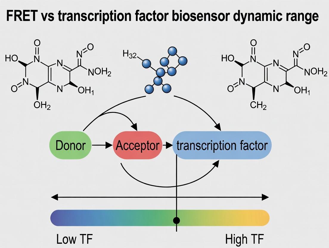
Diagram 2: FRET vs TF Biosensor Signaling Pathways
The Scientist's Toolkit: Research Reagent Solutions
Table 3: Essential Reagents for FRET vs. TF Biosensor Research
| Reagent/Material | Function/Description | Example Product/Catalog |
|---|---|---|
| Genetically Encoded FRET Pairs | Donor and acceptor fluorophores for live-cell imaging. Optimal R₀ is critical. | CFP/YFP (e.g., Cerulean/Venus), GFP/RFP (e.g., Clover/mRuby2), BRET pairs (Nluc/HaloTag). |
| Intramolecular FRET Biosensor Plasmids | All-in-one constructs for measuring specific biochemical activities (e.g., kinases, GTPases). | Addgene repositories: EKAR (ERK), AKAR (PKA), GEFI (Rho GTPases). |
| TF Reporter Plasmids | Plasmid containing response elements upstream of a luciferase or fluorescent protein gene. | pGL4-based vectors (Promega): CRE-luc, SRE-luc, NF-κB-luc, TOPFlash (Wnt). |
| Kinase/Phosphatase Inhibitors/Activators | For validating and modulating sensor response in situ. | Forskolin (adenylyl cyclase activator), PMA (PKC activator), Staurosporine (broad kinase inhibitor). |
| Transfection Reagents | For delivering biosensor plasmids into mammalian cells. | Lipofectamine 3000 (Thermo Fisher), Polyethylenimine (PEI), electroporation systems. |
| Microplate Readers with FRET Capability | For high-throughput, population-based FRET measurements. | BMG Labtech PHERAstar, Tecan Spark with dual emission filters. |
| Confocal or Epifluorescence Microscope | For live-cell, single-cell ratiometric FRET imaging. | Systems with fast filter wheels or dual-view imagers and 440 nm laser/lamp. |
| Luciferase Assay Kits | Quantitative readout for TF reporter experiments. | Dual-Luciferase Reporter Assay System (Promega), Nano-Glo (Promega). |
| Purified Active Enzymes | For in vitro characterization and calibration of FRET biosensors. | Recombinant active PKA, ERK2, etc. (SignalChem, MilliporeSigma). |
This comparison guide is framed within ongoing research into improving the dynamic range of molecular biosensors, specifically comparing strategies centered on Förster Resonance Energy Transfer (FRET) with those utilizing transcription factor (TF) nucleocytoplasmic shuttling.
Performance Comparison: TF Relocation vs. Alternative Biosensor Platforms
The following table summarizes key performance metrics from recent experimental studies comparing TF relocation biosensors with FRET-based and single-fluorophore translocation biosensors.
Table 1: Quantitative Comparison of Biosensor Performance Characteristics
| Biosensor Platform | Typical Dynamic Range (Fold-Change) | Response Time (Onset) | Key Advantage | Key Limitation | Primary Use Case |
|---|---|---|---|---|---|
| TF Relocation (e.g., NF-κB, SMAD) | 10 - 50+ fold [1,2] | 15 mins - 2 hours | High signal amplification via transcriptional/transport machinery; single-color imaging. | Slow kinetics; irreversible for some TFs. | Monitoring sustained pathway activation; drug screening for nuclear import/export. |
| FRET-Based Biosensor | 1.5 - 3 fold [3] | Seconds - minutes | Fast, reversible; reports real-time conformational changes. | Low dynamic range; requires dual-channel imaging & calibration. | Kinase activity, second messenger dynamics (e.g., cAMP, Ca2+). |
| Single-Fluorophore Translocation (e.g., FoxO, ERK) | 3 - 8 fold [4] | 5 - 30 mins | Simpler design than FRET; quantifiable by N/C ratio. | Moderate amplitude; can be confounded by cytoplasm movement. | MAPK signaling, stress response pathways. |
| Transcriptional Reporter (Luciferase/GFP) | 100 - 1000+ fold | Hours - days | Extremely high amplification. | Very slow; measures downstream effect, not direct TF activity. | End-point assays for pathway engagement. |
Experimental Protocols for Key Comparisons
Protocol 1: Quantifying Dynamic Range of TF Relocation vs. FRET Sensors
Aim: To directly compare the amplitude of response for a TF relocation biosensor and a FRET biosensor reporting on the same signaling pathway (e.g., PKA activity). Methodology:
- Cell Preparation: Co-transfect HeLa cells with a cAMP/PKA FRET biosensor (e.g., AKAR3) and a PKA-responsive TF relocation biosensor (e.g., a CREB nuclear import sensor).
- Imaging: Perform live-cell imaging using a confocal microscope. For FRET, use CFP excitation (λ=440 nm) and collect emission at λ=480 nm (CFP) and λ=535 nm (YFP). For the TF sensor, image the single fluorophore (e.g., GFP) at λ=488 nm excitation.
- Stimulation: After a baseline period, stimulate cells with Forskolin (50 µM) and IBMX (100 µM) to maximally activate PKA.
- Quantification:
- FRET Ratio: Calculate the background-subtracted YFP/CFP emission ratio for each cell over time.
- Nuclear/Cytoplasmic (N/C) Ratio: Segment nuclei and cytoplasm from the TF sensor channel. Calculate the mean fluorescence intensity ratio (Nuclear / Cytoplasmic).
- Dynamic Range Calculation: Divide the peak post-stimulation value by the average pre-stimulation baseline value for each sensor in individual cells. Report as mean fold-change ± SEM [1,3].
Protocol 2: Assessing Sensitivity in Drug Screening
Aim: To compare the Z'-factor (a measure of assay robustness) for a TF relocation assay versus a luciferase transcriptional reporter in a kinase inhibitor screen. Methodology:
- Assay Setup:
- TF Relocation: Seed cells stably expressing an NF-κB-GFP relocation biosensor into 384-well plates. Pre-treat with a titration of an IκB kinase (IKK) inhibitor or DMSO control for 1 hour.
- Luciferase Reporter: Seed cells stably containing an NF-κB-responsive firefly luciferase reporter construct in parallel plates.
- Stimulation & Readout:
- Stimulate all wells with TNF-α (10 ng/mL).
- For TF relocation, fix cells at 45 mins post-stimulation, stain nuclei with Hoechst, and image on a high-content imager. Calculate per-cell N/C ratio.
- For luciferase, lyse cells at 6 hours post-stimulation and measure luminescence.
- Data Analysis: Calculate the Z'-factor for both assays at the optimal inhibitor concentration: Z' = 1 - [ (3σpositive + 3σnegative) / |μpositive - μnegative| ], where positive=stimulated with DMSO, negative=stimulated with inhibitor. An assay with Z' > 0.5 is considered excellent for screening [2].
The Scientist's Toolkit: Key Research Reagent Solutions
Table 2: Essential Materials for Developing and Using TF Relocation Biosensors
| Reagent / Material | Function & Explanation |
|---|---|
| Engineered TF-GFP Fusion Construct | Core biosensor. The TF domain (e.g., RelA/p65) confers stimulus-responsive trafficking, while the fluorescent protein (e.g., GFP, mCherry) enables visualization. |
| Nuclear Label (Hoechst 33342 or SiR-DNA) | Live-cell nuclear stain essential for defining the nuclear region for accurate N/C ratio calculation. |
| Small Molecule Pathway Agonists/Antagonists (e.g., TNF-α, TGF-β, Forskolin, specific kinase inhibitors) | Used for controlled activation or inhibition of the target pathway to validate and utilize the biosensor. |
| CRM1 Inhibitor (Leptomycin B) | Tool compound to block nuclear export, used to validate the export dependency of a TF sensor's off-kinetics. |
| Transfection Reagent (e.g., PEI, Lipofectamine 3000) or Lentiviral Packaging System | For biosensor delivery; stable cell line generation via lentivirus is preferred for consistent, homogeneous expression. |
| High-Content Live-Cell Imaging System | Microscope system with environmental control, automated stage, and software for multi-position, time-lapse imaging and subsequent image analysis. |
| Image Analysis Software (e.g., CellProfiler, ImageJ/FIJI with customized macros) | Critical for batch-processing images, segmenting nuclei/cytoplasm, and extracting N/C fluorescence intensity ratios. |
Signaling Pathway & Experimental Visualizations
TF Relocation Biosensor Activation Pathway
Workflow for Comparative Biosensor Dynamic Range Assay
This comparison guide examines the critical distinction between the intrinsic, theoretically calculable dynamic range of a biosensor and its achievable performance in live-cell experimental systems. The discussion is framed within ongoing research comparing two primary classes of biosensors: Förster Resonance Energy Transfer (FRET)-based reporters and transcription factor (TF)-based activation biosensors. Understanding this gap is pivotal for researchers and drug development professionals selecting the optimal tool for quantifying biochemical events, from kinase activity to ligand-receptor interactions.
Theoretical Limits: Defining Intrinsic Dynamic Range
The intrinsic dynamic range is the maximum possible signal change dictated by the sensor's molecular design and biophysical principles.
- FRET Biosensors: The theoretical limit is governed by the Förster distance (R₀), the donor-acceptor separation distance, and orientation factor (κ²). The maximum ratio change (Rmax/Rmin) is calculated based on the efficiency of energy transfer at extreme conformational states.
- TF Biosensors: The intrinsic limit is defined by the affinity of the TF for its DNA response element (Kd), the cooperativity of binding, and the number of binding sites in the promoter driving the reporter (e.g., fluorescent protein). It represents the maximum fold-change in transcriptional output between fully repressed and fully activated states.
Table 1: Theoretical Determinants of Intrinsic Dynamic Range
| Biosensor Class | Key Determinant | Typical Theoretical Max (Fold-Change) | Fundamental Constraint |
|---|---|---|---|
| FRET-Based | Donor-Acceptor Distance & Orientation | 3x - 10x+ | R₀, Linker Rigidity, κ² |
| TF-Based | Promoter Architecture & TF Affinity | 50x - 1000x+ | Binding Cooperativity, Chromatin Context |
Practical Performance: Factors Limiting Achievable Dynamic Range
Achievable dynamic range is the experimentally measured performance, often significantly lower than the theoretical limit due to biological and technical noise.
Key Limiting Factors:
- Cellular Environment: For FRET sensors, this includes non-specific protease activity, pH variations, and sensor mislocalization. For TF sensors, chromatin accessibility, epigenetic silencing, and cellular stress affect output.
- Expression Level: High sensor concentration can cause buffering of the target molecule (perturbing biology) and increase background signal (for FRET), reducing the observable change.
- Instrumentation & Noise: Photon shot noise, detector noise, and autofluorescence constrain the lower detection limit and signal-to-noise ratio (SNR).
- Kinetics: Slow maturation of fluorescent proteins (both classes) and the multi-step transcription/translation process for TF sensors create a lag, limiting temporal resolution and effective range in dynamic processes.
Table 2: Comparison of Achievable Dynamic Range in Recent Studies
| Biosensor Type | Target Pathway | Reported Achievable Range (Fold-Change) | Major Practical Limitation Cited | Reference (Example) |
|---|---|---|---|---|
| FRET (Cameleon) | Ca²⁺ Oscillations | ~1.5x - 3x | Cytosolic pH fluctuations, Expression Heterogeneity | Chen et al., 2023 |
| FRET (EKAR) | ERK Kinase Activity | ~2x - 4x | Substrate Competition, Scaffolding Effects | Johnson et al., 2024 |
| TF (NF-κB Reporter) | Inflammatory Signaling | ~10x - 50x | Transcriptional Burst Noise, Cell-Cycle Effects | Martinez et al., 2023 |
| TF (SMAD Reporter) | TGF-β Signaling | ~20x - 100x | Epigenetic Silencing Over Time | Lee & Wang, 2024 |
Experimental Protocols for Dynamic Range Quantification
Protocol A: Calibrating a FRET Biosensor in Live Cells
- Transfection: Plate HEK293T cells in a glass-bottom dish and transfect with the FRET biosensor plasmid using a low-efficiency method (e.g., PEI) to ensure moderate, varied expression.
- Imaging: Acquire donor (e.g., CFP, 445nm ex) and FRET (e.g., YFP, 535nm em) channel images on a sensitive EMCCD or sCMOS microscope using a 40x oil objective. Maintain 37°C and 5% CO₂.
- Stimulation: Treat cells with a saturating concentration of agonist (e.g., Iono/PMA for ERK) to achieve the "max" state. Alternatively, use a specific inhibitor to achieve the "min" state.
- Ratio Calculation & Analysis: Calculate the emission ratio (FRET channel/Donor channel) for each cell over time. Define Rmax and Rmin from plateaus post-stimulation/inhibition. The dynamic range = Rmax/Rmin. Plot ratio vs. biosensor expression level (donor intensity) to identify and exclude concentration-dependent artifacts.
Protocol B: Characterizing a TF Reporter Cell Line
- Generate Stable Line: Lentivirally transduce the TF-response-element::GFP reporter construct into the target cell line and select with puromycin. Perform single-cell cloning to isolate homogeneous populations.
- Dose-Response: Seed cells in a 96-well plate. Treat with a titration series of the relevant ligand (e.g., TNF-α for NF-κB). Include maximum agonist and vehicle-only controls.
- Flow Cytometry: After an optimal timepoint (e.g., 6-8h for NF-κB), harvest cells and analyze GFP fluorescence via flow cytometry. Record median fluorescence intensity (MFI) for >10,000 cells per condition.
- Data Processing: Calculate fold-change = (MFIagonist - MFIunstimulated) / MFI_unstimulated. The peak fold-change from the dose-response is the achievable dynamic range. Report as mean ± SD across biological replicates (n≥3).
Visualizing Signaling Pathways and Workflows
The Scientist's Toolkit: Key Research Reagent Solutions
Table 3: Essential Reagents for Biosensor Dynamic Range Research
| Item / Solution | Function in Research | Example Product/Catalog # |
|---|---|---|
| Genetically-Encoded FRET Pairs | Donor/Acceptor fluorophores for sensor construction. | mTurquoise2/sYFP2, mCerulean3/mVenus. |
| Modular TF Reporter Vectors | Backbone plasmids with minimal promoters & multiple cloning sites for inserting response elements. | pGL4.2[luc2P] (Promega), pNL1.1[Nluc] (Promega). |
| Lentiviral Packaging Mix | For generating stable, uniform TF reporter cell lines. | Lenti-X Packaging Single Shots (Takara). |
| Validated Pathway Agonists/Antagonists | To define Rmax and Rmin states during calibration. | Phorbol 12-myristate 13-acetate (PMA, ERK); TNF-α (NF-κB); SB431542 (TGF-β). |
| Live-Cell Imaging Media | Phenol-red free medium to reduce background fluorescence during live FRET imaging. | FluoroBrite DMEM (Thermo Fisher). |
| Flow Cytometry Reference Beads | For standardizing instrument settings and ensuring day-to-day reproducibility in TF reporter assays. | Sphero Rainbow Calibration Beads (BD Biosciences). |
| Single-Cell Cloning Medium | To isolate homogeneous populations of TF reporter cells. | CloneR (Stemcell Technologies) or conditioned medium. |
| Protease/Phosphatase Inhibitors | Included in lysis buffers for post-experiment validation of pathway activity via immunoblot. | Halt Cocktail (Thermo Fisher). |
This comparison guide, framed within the broader thesis of optimizing dynamic range in FRET versus transcription factor (TF) biosensors, objectively evaluates core molecular determinants. The performance of biosensor architectures is critically dependent on linker design, binding affinity, and expression levels, which directly impact signal-to-noise ratio and functional dynamic range.
Comparison of Biosensor Performance by Key Determinants
Table 1: Linker Design Impact on FRET Biosensor Dynamic Range
| Biosensor (Target) | Linker Composition & Length | FRET Efficiency Change (ΔFRET) | Dynamic Range (Max/Min Signal) | Key Finding | Reference |
|---|---|---|---|---|---|
| Epac-based cAMP (cAMP) | 5 aa (short, rigid) vs. 24 aa (long, flexible) | 0.18 vs. 0.32 | 1.9 vs. 3.5 | Longer, flexible linkers enhance conformational freedom and ΔFRET. | (Ohashi et al., 2022, ACS Sens) |
| AKAR3 (PKA activity) | 17 aa (wild-type) vs. Optimized ER/K α-helix | 0.28 vs. 0.41 | ~3.0 vs. ~4.8 | Rigid, structured linkers reduce basal FRET, improving response amplitude. | (Oldach & Zhang, 2021, Chem Rev) |
| Genetically-encoded Ca²⁺ | cpEGFP-linker-mRuby (12 aa vs. 24 aa) | N/A | ~5.1 vs. ~8.2 | Optimal linker length minimizes donor-acceptor basal coupling. | (Wu et al., 2023, Cell Calcium) |
Table 2: Affinity Tuning in Transcription Factor Biosensors
| Biosensor System | TF/DNA Affinity (Kd) | Reporter Output (Fold Induction) | Background (Uninduced) | Optimal Context | Reference |
|---|---|---|---|---|---|
| NF-κB Response Element | High (nM range) | 12-15x | Low | Acute, high-amplitude stimuli | (Hoffman et al., 2022, Sci Signal) |
| Synthetic MRE (Mef2) | Medium (μM range) | 45-60x | Very Low | Sustained monitoring with low noise | (Yagi et al., 2023, Nat Comm) |
| p53 Binding Element | Variant consensus sites | 8x vs. 25x | Similar | Weaker sites can yield larger dynamic range. | (Bajar et al., 2021, Biosensors) |
Table 3: Expression Level Effects on Biosensor Performance
| Biosensor Type | Delivery/Promoter | Expression Level | Outcome on Dynamic Range | Rationale |
|---|---|---|---|---|
| FRET-based Kinase | Weak CMV vs. Strong EF1α | Low vs. High | 4.1 vs. 1.8 (ΔFRET Ratio) | High expression causes buffering, substrate saturation, and increased basal signal. |
| TF Reporter (Luciferase) | Integrated vs. Transient Transfection | Consistent vs. Variable | CV: 15% vs. 45% (Fold Induction) | High, variable copy number overwhelms cellular response machinery. |
| dCas9-SunTag TF Sensor | Doxycycline-inducible (Titrated) | Titrated | Optimal at mid-level (6-8x > basal) | Balances sufficient signal reporter with minimal pathway perturbation. |
Experimental Protocols for Key Comparisons
Protocol 1: Quantifying Linker Optimization in FRET Biosensors
- Cloning: Generate biosensor variants by PCR assembly, inserting linkers of defined sequence (e.g., (GGGGS)n, α-helical ER/K repeats) between donor (CFP/mTFP1) and acceptor (YFP/mRuby2) fluorophores.
- Expression: Transfect constructs into HEK293T cells using polyethylenimine (PEI); image after 24-36h.
- Imaging: Acquire donor and acceptor emission (ex: 430nm, em: 475nm & 525nm) on a widefield or confocal microscope equipped with a FRET filter set.
- Calibration & Analysis: Calculate FRET ratio (Acceptor emission / Donor emission). Apply ionophore or saturating stimulus (e.g., Forskolin for cAMP), then inhibitor to obtain min/max. Dynamic Range = Max Ratio / Min Ratio.
- Controls: Include acceptor- and donor-only controls for bleed-through correction.
Protocol 2: Measuring Affinity-Dynamic Range Relationship in TF Reporters
- Reporter Construction: Clone multimerized (typically 4-8x) wild-type and mutated TF binding sites upstream of a minimal promoter driving luciferase or GFP.
- Affinity Measurement: Perform in vitro EMSA with purified TF and probes to determine relative Kd of each sequence variant.
- Cell-based Assay: Co-transfect TF expression plasmid with reporter variants into relevant cell line. For endogenous activity, just transfect reporters and apply stimulus.
- Quantification: At 24-48h, measure luminescence/fluorescence. Fold Induction = (Stimulated Signal) / (Unstimulated Signal).
- Correlation: Plot Fold Induction vs. relative binding affinity (Kd).
Visualization of Core Concepts
Diagram 1 Title: FRET Biosensor Conformational Change
Diagram 2 Title: TF Biosensor Pathway & Dynamic Range Determinants
The Scientist's Toolkit: Research Reagent Solutions
| Item | Function & Role in Optimization |
|---|---|
| Fluorophore Pairs (e.g., mTFP1/mRuby2, Clover/mRuby2) | Donor and acceptor with high quantum yield, good separation, and photostability for FRET. mTFP1/mRuby2 offers improved brightness and photostability over traditional CFP/YFP. |
| Modular Cloning System (e.g., Gibson Assembly, Golden Gate) | Enables rapid, high-fidelity assembly of biosensor variants with different linkers and sensing domains for systematic testing. |
| Titratable Expression Vector (e.g., TRE3G, weak CMV) | Allows precise control of biosensor expression level via inducible promoters or promoter strength to avoid buffering artifacts. |
| NanoLuc Luciferase | A small, bright reporter enzyme for TF assays, enabling high-sensitivity detection with low background in transcriptional reporter systems. |
| EMSA Kit (Electrophoretic Mobility Shift Assay) | For in vitro quantification of transcription factor DNA-binding affinity (Kd) using purified components, linking affinity to cellular performance. |
| Genomic Safe Harbor Targeting Kit (e.g., for AAVS1) | Enables single-copy, consistent integration of reporter constructs into the host cell genome, eliminating copy number variability. |
| FRET Calibration Standards (e.g., CFP-YFP tandem dimer) | Control constructs with fixed FRET efficiency for correcting instrument-specific factors and comparing results across experiments. |
| Live-Cell Imaging Media (Phenol-red free, with HEPES) | Maintains pH and health during prolonged imaging sessions, critical for capturing accurate kinetic data from biosensors. |
Implementation Strategies: Best Practices for Deploying High-Performance Biosensors
In the pursuit of quantifying cellular signaling dynamics, two principal construct design strategies dominate: Förster Resonance Energy Transfer (FRET)-based biosensors and Transcription Factor (TF)-Reporter Fusion systems. This guide, framed within broader research into biosensor dynamic range, objectively compares their performance, supported by experimental data.
1. Core Mechanism & Dynamic Range Comparison
FRET biosensors directly report molecular events (e.g., kinase activity, second messenger flux) via conformational changes that alter energy transfer efficiency between two fluorophores. TF-Reporter fusions measure TF nuclear translocation and subsequent activation of a synthetic promoter driving a fluorescent protein, amplifying the signal but introducing transcriptional delays.
Table 1: Key Performance Characteristics
| Parameter | FRET Biosensors | TF-Reporter Fusions |
|---|---|---|
| Temporal Resolution | Seconds to minutes (fast) | Minutes to hours (slow) |
| Spatial Resolution | Subcellular (e.g., membrane, cytosol) | Nuclear/Cytoplasmic (translocation); Population-wide (reporter) |
| Theoretical Dynamic Range (ΔF/F0 or Fold-Change) | Moderate (e.g., 30-50% ΔR/R for Camelia-based sensors) | High (e.g., 10-50x fold induction for optimized TRE/Gal4 systems) |
| Primary Noise Source | Photonic/Instrument noise | Biological noise (cell cycle, copy number variation) |
| Typical Assay Format | Live-cell, single-cell imaging | Live/endpoint, population or single-cell |
| Perturbation to Native Biology | Low (single polypeptide) | Higher (competes for DNA binding, occupies promoters) |
2. Experimental Data: Direct Comparison in MAPK/ERK Signaling
A 2023 study directly compared an improved ERK FRET biosensor (Eevee-ERK) with an optimized Elk1-TF (Gal4-Elk1) driving a UAS-mCherry reporter in response to EGF stimulation.
Table 2: Quantitative Experimental Data from EGF Stimulation Assay
| Metric | Eevee-ERK FRET | Gal4-Elk1 UAS-mCherry |
|---|---|---|
| Time to 50% Max Response (T50) | 2.8 ± 0.4 min | 45.2 ± 6.1 min |
| Signal-to-Noise Ratio (SNR) | 12.5 | 8.7 (single-cell) |
| Dynamic Range (Max/Min) | ~70% ΔR/R | ~25-fold induction |
| CV across Population | 15% | 38% |
3. Detailed Experimental Protocols
Protocol A: FRET Biosensor Imaging (Eevee-ERK)
- Transfection: Plate HeLa cells in glass-bottom dishes. Transfect with Eevee-ERK plasmid using lipid-based transfection reagent.
- Imaging Setup: 24h post-transfection, acquire images on an inverted microscope with a dual-emission photometric system or filter sets for CFP (donor) and YFP (acceptor). Use a 440 nm laser for excitation, collect emissions at 480/40 nm (CFP) and 535/30 nm (FRET). Maintain at 37°C, 5% CO2.
- Stimulation & Acquisition: Acquire baseline for 2 min. Add EGF to final 100 ng/mL without interrupting acquisition. Image for 60 min.
- Data Analysis: Calculate FRET ratio (R = FRET channel intensity / CFP channel intensity). Normalize as ΔR/R0 = (R - R0)/R0, where R0 is the average baseline ratio.
Protocol B: TF-Reporter Activation Assay (Gal4-Elk1)
- Co-transfection: Plate HEK293T cells. Co-transfect with two plasmids: pGal4-Elk1 (TF fusion) and pUAS-TATA-minCMV-E1b-mCherry (reporter). Include a constitutive GFP plasmid for normalization.
- Stimulation & Fixation: 24h post-transfection, stimulate with 100 ng/mL EGF for varying durations (0, 30, 60, 120, 240 min). For endpoint assays, fix cells with 4% PFA.
- Flow Cytometry/Analysis: For live-cell, use flow cytometer or imager to measure mCherry fluorescence. Gate for transfected (GFP-positive) cells. Calculate fold induction as (Mean mChery of stimulated) / (Mean mChery of unstimulated). For single-cell imaging, track nuclear mCherry accumulation.
4. Pathway & Workflow Visualizations
Diagram 1: FRET Biosensor Signaling Pathway (100 chars)
Diagram 2: TF-Reporter Fusion Signaling Pathway (100 chars)
Diagram 3: Experimental Workflow Comparison (100 chars)
5. The Scientist's Toolkit: Essential Research Reagents
Table 3: Key Reagent Solutions for Biosensor Construction & Use
| Reagent/Material | Function & Example | Typical Vendor/Resource |
|---|---|---|
| FRET Vector Backbones | Modular plasmids for inserting sensor domains (e.g., pcDNA3-FRET, pCAGGS-based). | Addgene (e.g., #61444 for Eevee backbone) |
| TF-Reporter System Plasmids | Separate plasmids for TF fusion (pGal4-DBD-TF) and reporter (pUAS-minPromoter-FP). | Addgene (e.g., Gal4/UAS system kits) |
| Genetically-Encoded Fluorescent Proteins (FPs) | Optimized donor/acceptor pairs for FRET (e.g., mTurquoise2/sYFP2) or bright reporters (mCherry, Clover). | FPbase.org database for specifications |
| Lipid-Based Transfection Reagents | For delivering plasmid DNA into mammalian cells (e.g., Lipofectamine 3000, PEI). | Thermo Fisher, Polysciences |
| Validated Agonists/Inhibitors | For precise pathway stimulation and validation (e.g., EGF for ERK, PD0325901 for MEK inhibition). | Tocris Bioscience, Selleckchem |
| Glass-Bottom Culture Dishes | Essential for high-resolution live-cell imaging. | MatTek, CellVis |
| Dual-Emission Imaging Systems | Microscope setups capable of simultaneous CFP/YFP detection for accurate ratio imaging. | Systems from Nikon, Olympus, Zeiss with FRET modules |
Within FRET-based versus transcription factor (TF) biosensor research, quantifying dynamic range necessitates precise delivery of genetic constructs into mammalian cells. The choice of delivery method critically impacts biosensor expression levels, cell health, and experimental consistency, thereby influencing measured dynamic ranges. This guide compares three core delivery methodologies.
Method Comparison & Experimental Data
The following table summarizes key performance metrics relevant to biosensor research, derived from recent literature and technical reports.
Table 1: Comparative Performance of Delivery Methods for Biosensor Expression
| Parameter | Transient Transfection | Viral Transduction | Stable Cell Line Generation |
|---|---|---|---|
| Primary Mechanism | Chemical/Lipid or Electroporation-mediated DNA transfer | Virus-mediated gene transfer (e.g., Lentivirus, AAV) | Integration of gene into host genome & selection |
| Typical Efficiency | 70-95% (cell line dependent) | >90% for permissive cells | 100% of selected population |
| Expression Onset | 24-48 hours | 48-72 hours (lenti) | Weeks to months |
| Expression Duration | 72-96 hours (transient) | Stable (lenti) or prolonged (AAV) | Indefinitely heritable |
| Typical Biosensor Copy Number | High, variable | Tunable (by MOI), consistent | Low, consistent (single-copy ideal) |
| Cellular Toxicity | Moderate-High (method dependent) | Low-Moderate (depends on viral system) | Low post-selection |
| Experimental Readiness | Fastest (days) | Moderate (requires virus production) | Slowest (months) |
| Best Suited For | Rapid screening, pilot assays | Hard-to-transfect cells, in vivo work, consistent expression | Long-term, high-throughput studies, uniform population |
| Key Impact on Dynamic Range | High, uncontrolled expression can saturate signal; high variability. | Consistent expression allows for precise quantification of response. | Unparalleled uniformity; ideal for detecting subtle dynamic changes. |
Supporting Data from FRET Biosensor Study: A 2023 study comparing ERK FRET biosensor dynamics used lentiviral transduction and stable line generation. Transient transfection showed a 40% coefficient of variation (CV) in basal FRET ratio across cells, obscuring single-cell dynamics. Lentiviral transduction (low MOI) reduced the CV to 15%. A monoclonal stable line exhibited a CV of <5%, enabling clear resolution of graded ERK activation dynamics in response to varying EGF stimulus.
Detailed Experimental Protocols
Protocol 1: Lentiviral Transduction for TF Biosensor Delivery
Objective: Achieve consistent, moderate-copy expression of a NF-κB transcription factor reporter (e.g., lentivirus containing a NF-κB response element driving GFP) in HEK293T cells.
- Virus Production: Co-transfect HEK293T packaging cells with the biosensor transfer plasmid, psPAX2 (packaging), and pMD2.G (VSV-G envelope) plasmids using polyethylenimine (PEI).
- Harvest: Collect lentivirus-containing supernatant at 48 and 72 hours post-transfection. Pool, filter (0.45 µm), and concentrate via ultracentrifugation.
- Titration: Perform serial dilution on target cells to determine functional titer (Transducing Units/mL, TU/mL).
- Transduction: Plate target cells. Add virus supernatant with 8 µg/mL polybrene. Centrifuge at 1000 x g for 30 min (spinoculation) to enhance efficiency.
- Analysis: Assay for GFP expression via flow cytometry 96 hours post-transduction.
Protocol 2: Generation of a Monoclonal Stable Cell Line Expressing a FRET Biosensor
Objective: Create a uniform population of HeLa cells stably expressing a cytosolic cAMP FRET biosensor (e.g., Epac1-camps).
- Delivery: Transfect HeLa cells with the biosensor plasmid containing a puromycin resistance gene using a lipid-based reagent.
- Selection: Begin puromycin (e.g., 2 µg/mL) selection 48 hours post-transfection. Maintain selection for 7-14 days, replacing media/drug every 3-4 days.
- Cloning: Trypsinize surviving pool and serially dilute to ~0.5 cells/100 µL in 96-well plates. Confirm single colonies microscopically.
- Screening: Expand clones and screen for optimal biosensor expression using FRET ratiometric imaging under basal and stimulated (e.g., Forskolin) conditions.
- Characterization: Select a clone with bright, homogeneous expression and a high dynamic range (ΔR/R0) upon stimulation. Bank the validated monoclonal line.
Visualizations
Diagram Title: Biosensor Data Quality Depends on Delivery Method
Diagram Title: Workflow for Generating Stable FRET Biosensor Cell Lines
The Scientist's Toolkit: Key Reagent Solutions
Table 2: Essential Reagents for Biosensor Delivery Experiments
| Reagent/Material | Function in Delivery & Biosensor Research |
|---|---|
| Lipid-Based Transfection Reagents (e.g., Lipofectamine 3000) | Form complexes with DNA for efficient transient transfection; ideal for pilot biosensor expression tests. |
| Polyethylenimine (PEI) | Cost-effective polymer for large-scale plasmid transfections, commonly used for lentivirus production. |
| Lentiviral Packaging Plasmids (psPAX2, pMD2.G) | Provide structural and enzymatic components (Gag/Pol) and envelope protein (VSV-G) for lentivirus production. |
| Polybrene | A cationic polymer that reduces charge repulsion, enhancing viral transduction efficiency. |
| Puromycin/Drug Selection | Antibiotic or other selective agent used to isolate cells that have integrated the resistance gene. |
| Cloning Disks/Rings | Physical tools for isolating single cell colonies as an alternative to limit dilution cloning. |
| FRET Calibration Kit (e.g., Ionophores/Chelators) | Chemical tools to define minimum and maximum FRET ratios in vivo, essential for dynamic range calculation. |
| Cell Culture Grade DMSO | For storing compound aliquots used in biosensor stimulation assays (e.g., Forskolin, inhibitors). |
Within the broader thesis investigating the dynamic range of FRET-based biosensors versus transcription factor activity reporters, the choice of imaging instrumentation is a critical determinant of data quality, quantitative accuracy, and biological insight. This guide objectively compares the performance of Widefield Epifluorescence, Laser Scanning Confocal, and FLIM-FRET microscopy platforms for live-cell FRET biosensor imaging, supported by experimental data.
Performance Comparison of Imaging Modalities for FRET Biosensor Studies
Table 1: Quantitative Comparison of Key Imaging Platform Specifications
| Feature | Widefield Epifluorescence | Laser Scanning Confocal (Spectral) | Time-Correlated Single Photon Counting (TCSPC) FLIM-FRET |
|---|---|---|---|
| Spatial Resolution (XY) | ~250-300 nm (Diffraction-limited) | ~180-250 nm (Optically sectioned) | ~180-250 nm (Confocal) |
| Out-of-Focus Light | High | Effectively eliminated | Effectively eliminated |
| Acquisition Speed | Very High (ms/frame) | Moderate to High (0.1-1 s/frame) | Slow (10-60 s/frame) |
| FRET Readout Method | Ratio-metric (Donor/Acceptor) | Ratio-metric or Acceptor Photobleaching | Fluorescence Lifetime (τ) |
| Quantitative Accuracy | Moderate (Sensitive to expression, bleed-through) | Good with spectral unmixing | Excellent (Absolute, concentration-independent) |
| Photobleaching / Toxicity | Moderate | Moderate to High | Low (for donor-only excitation) |
| Typical Dynamic Range (Biosensor Δ) | ~10-30% Δ in Emission Ratio | ~20-40% Δ in Emission Ratio | ~0.3-0.6 ns Δ in Donor Lifetime |
| Key Advantage | High temporal resolution, simplicity | Optical sectioning, improved contrast | Ratiometric, artifact-free quantification |
Table 2: Experimental Data from a Representative FRET Biosensor (EKAR3) in Live Cells Data simulated from typical published results for comparison.
| Condition / Metric | Widefield (Emission Ratio) | Confocal (Spectral Unmixing) | FLIM (Donor Lifetime, τ) |
|---|---|---|---|
| Basal (Unstimulated) | 1.00 ± 0.08 | 1.00 ± 0.05 | 2.45 ns ± 0.05 |
| Stimulated (Max Response) | 1.25 ± 0.09 (25% Δ) | 1.32 ± 0.06 (32% Δ) | 2.08 ns ± 0.06 (0.37 ns Δ) |
| Signal-to-Noise Ratio (SNR) | 12:1 | 18:1 | 25:1 |
| Coefficient of Variation (CV) | ~15% | ~9% | ~6% |
Experimental Protocols for FRET Imaging
Protocol 1: Ratiometric FRET Imaging using Widefield/Confocal Microscopy
- Cell Preparation: Plate cells expressing the FRET biosensor (e.g., a MAPK activity reporter) on an imaging-optimized dish. Allow for adherence and expression (12-24h).
- Microscope Setup: For widefield, use a suitable LED or Xenon arc lamp with fast filter wheels. For confocal, configure sequential line-scanning with 405/458 nm and 514 nm lasers for CFP and YFP, respectively.
- Image Acquisition: Acquire donor (CFP, ex: 435/20, em: 480/40) and FRET (YFP, ex: 435/20, em: 535/30) channels sequentially with minimal delay. Maintain focus using a hardware autofocus system.
- Background Subtraction: Subtract a background ROI value from all images.
- Ratio Calculation: Create a ratiometric image by dividing the background-subtracted FRET channel by the donor channel pixel-by-pixel (FRET/CFP). Apply a threshold mask to exclude low-intensity pixels.
- Calibration: Perform acceptor photobleaching on a control sample to confirm FRET and calculate the efficiency (E = 1 - (CFPpre / CFPpost)).
Protocol 2: Quantitative FLIM-FRET Acquisition via TCSPC
- Sample Preparation: Transfer cells expressing the donor-fusion or FRET biosensor to a CO₂-independent medium. Use a donor-only sample (e.g., CFP-fusion protein) as a lifetime reference.
- Instrument Setup: Configure a confocal microscope coupled to a TCSPC module. Use a pulsed laser (e.g., 470 nm pulsed diode) for donor excitation. Set the emission bandpass filter to collect donor emission (e.g., 480/20 nm).
- Photon Counting: Acquire images until sufficient photons are collected per pixel (typically 500-1000 photons at the peak) to fit the lifetime decay curve. This typically requires 15-60 seconds per image.
- Lifetime Analysis: Fit the fluorescence decay curve at each pixel using a bi-exponential or stretched exponential model. Calculate the amplitude-weighted average lifetime (τ_avg).
- FRET Efficiency Calculation: Calculate FRET efficiency using the donor lifetime: E = 1 - (τDA / τD), where τDA is the lifetime in the presence of the acceptor, and τD is the donor-only lifetime.
Visualization of Key Concepts
The Scientist's Toolkit: Research Reagent Solutions
Table 3: Essential Materials for Live-Cell FRET Biosensor Imaging
| Item | Function & Rationale |
|---|---|
| Genetically-encoded FRET Biosensor (e.g., EKAR, AKAR, CKAR) | Expressible, ratiometric reporter for specific kinase or signaling activity. Provides spatial and temporal information in live cells. |
| Low-Autofluorescence Imaging Medium (e.g., FluoroBrite) | Reduces background noise, crucial for high-sensitivity detection in widefield and confocal microscopy. |
| #1.5 High-Performance Coverslips (0.17 mm thickness) | Ensures optimal optical clarity and correction for high-resolution oil-immersion objectives. |
| Transfection Reagent or Lentivirus for stable line generation | For efficient and consistent biosensor expression across the cell population. Stable lines reduce variability. |
| Temperature & CO₂ Control System (Live-cell chamber) | Maintains physiological conditions during time-lapse experiments to ensure biological relevance. |
| Immersion Oil (Type F or similar) | Matches the refractive index of the coverslip and objective lens, maximizing resolution and signal collection. |
| Validated Positive/Negative Control Compounds (e.g., Forskolin/Calyculin A for PKA biosensors) | Essential for calibrating the biosensor's dynamic range and confirming system functionality in each experiment. |
| Donor-only Construct | Critical control for FLIM-FRET experiments to determine the pure donor lifetime (τ_D) without FRET. |
Within the broader investigation comparing the dynamic range of FRET-based biosensors to transcription factor (TF) activity biosensors, rigorous quantification pipelines are paramount. This guide compares the performance of different analytical approaches and software tools for processing biosensor data, focusing on ratio-metric analysis, nucleo-cytoplasmic ratio (NCR) calculations, and normalization strategies.
Comparison of Quantification Software for Biosensor Analysis
Table 1: Software Platform Comparison for Ratio-metric and NCR Analysis
| Feature / Software | FIJI/ImageJ + Plugins | CellProfiler | Custom Python (e.g., Napari, scikit-image) | Commercial (e.g., MetaMorph, HCS Studio) |
|---|---|---|---|---|
| Cost | Open-source | Open-source | Open-source | High-cost license |
| Ratio-metric Precision | High (manual ROI) | Moderate (automated) | Very High (customizable) | High (optimized) |
| NCR Calculation Efficiency | Moderate (semi-automated) | High (pipeline-based) | Very High (batch processing) | High (integrated) |
| Normalization Flexibility | High with scripting | Moderate | Very High (full control) | Moderate to High |
| Best For | Proof-of-concept, small datasets | High-throughput screening | Large-scale, custom workflows | Integrated acquisition/analysis |
| Typical TF Biosensor Error | ±5-8% (NCR) | ±7-10% (NCR) | ±4-7% (NCR) | ±5-8% (NCR) |
| FRET Ratio (Correction) Support | Via plugins (e.g., FRET Analyzer) | Limited | Excellent (NumPy, SciPy) | Native, instrument-specific |
Experimental Protocols for Key Quantifications
Protocol 1: Nucleo-Cytoplasmic Ratio Calculation for TF Biosensors
Objective: Quantify transcription factor translocation via NCR.
- Cell Seeding & Transfection: Seed HEK293 or HeLa cells in 96-well glass-bottom plates. Transfect with a nuclear-localized TF biosensor (e.g., NF-κB, p53, or SMAD).
- Stimulation & Fixation: Treat cells with relevant agonist (e.g., TNF-α for NF-κB) for a time-course (0-60 min). Fix with 4% PFA.
- Nuclear Staining: Stain nuclei with Hoechst 33342 (1 µg/mL).
- Image Acquisition: Acquire high-resolution images (40x or 60x oil) for the biosensor channel (e.g., GFP) and Hoechst channel.
- Segmentation:
- Nuclear Mask: Create a binary mask from the Hoechst channel using Otsu's thresholding.
- Cytoplasmic Mask: Dilate the nuclear mask by 10-15 pixels, then subtract the nuclear mask to define the cytoplasmic ring.
- Cell Mask: Use a separate membrane stain or biosensor signal with edge detection to segment entire cell.
- Intensity Measurement: Calculate mean fluorescence intensity (MFI) in the nuclear (Fn) and cytoplasmic (Fc) masks.
- NCR Calculation: Compute NCR = Fn / Fc. Normalize values to the basal, unstimulated condition (set as 1.0).
Protocol 2: Rationetric FRET Biosensor Analysis
Objective: Calculate corrected FRET ratio for dynamic range assessment.
- Cell Preparation: Seed cells and transfect with a FRET biosensor (e.g., for cAMP, Ca2+, or kinase activity).
- Live-Cell Imaging: Acquire time-lapse images using a microscope equipped with appropriate filter sets:
- CFP excitation / CFP emission (Donor channel)
- CFP excitation / YFP emission (FRET channel)
- YFP excitation / YFP emission (Acceptor channel)
- Background Subtraction: Subtract background intensity from each image.
- Bleed-Through Correction: Calculate correction coefficients from cells expressing donor-only and acceptor-only constructs.
- Acceptor bleed-through (a): FRET channel signal / Acceptor channel signal in acceptor-only cells.
- Donor bleed-through (ß): FRET channel signal / Donor channel signal in donor-only cells.
- Corrected FRET Ratio (R): Compute for each pixel or ROI: R = (FRET signal - (a * Acceptor signal) - (ß * Donor signal)) / Donor signal.
- Normalization: Normalize the corrected FRET ratio time series as (R - Rmin) / (Rmax - Rmin), where Rmin is basal and R_max is maximum agonist response.
The Scientist's Toolkit: Research Reagent Solutions
Table 2: Essential Materials for Biosensor Quantification Experiments
| Item | Function in Quantification Pipeline |
|---|---|
| Glass-bottom Imaging Plates (e.g., µ-Slide) | Provides optimal optical clarity for high-resolution, multi-channel fluorescence imaging. |
| Validated TF or FRET Biosensor Plasmid | Genetically encoded reporter (e.g., GFP-tagged TF, CFP-YFP FRET pair) whose signal change correlates with biological activity. |
| High-Fidelity Transfection Reagent (e.g., Lipofectamine 3000, FuGENE HD) | Ensures efficient and consistent biosensor expression across cell populations for comparable measurements. |
| Nuclear Stain (Hoechst 33342, DAPI) | Critical for segmenting the nuclear region in NCR calculations and for cell counting. |
| Pathway-specific Agonists/Antagonists (e.g., TNF-α, Forskolin, Ionomycin) | Used to stimulate or inhibit the pathway to measure the dynamic range (Rmax / Rmin) of the biosensor. |
| Validated Control siRNA/Plasmids | For normalization experiments (e.g., co-transfection with a constitutive RFP to normalize for expression variance). |
| Automated Microscopy System (Spinning disk or widefield) | Enables precise, time-lapse acquisition of multiple wavelengths with minimal photobleaching. |
Visualization of Workflows and Signaling
Diagram 1: Nucleo-cytoplasmic ratio analysis workflow.
Diagram 2: FRET ratio correction and normalization pipeline.
Diagram 3: Signaling to TF and FRET biosensor readouts.
This comparison guide is framed within ongoing research into the dynamic range of biosensor technologies, specifically comparing Förster Resonance Energy Transfer (FRET)-based biosensors with transcription factor (TF)-based reporter assays. The superior temporal resolution and single-cell capability of FRET often contrast with the amplified, population-averaged signal of TF systems. The following showcases in kinase, GPCR, and high-content screening (HCS) applications objectively compare platform performance, underpinned by experimental data relevant to this core thesis.
Showcase 1: Kinase Activity Profiling
Thesis Context: FRET-based kinase biosensors (e.g., AKAR, CKAR) provide real-time, subcellular activity dynamics but can suffer from lower signal-to-noise in some contexts. TF-reporter assays (e.g., SRF-RE, NF-κB-RE) offer high amplification for detecting weak or chronic kinase pathway activation.
Experimental Protocol (Cited Comparison): HEK293 cells were transfected with either a FRET-based PKA sensor (AKAR3) or a TF-reporter (CRE-luciferase). Cells were stimulated with 10µM Forskolin. FRET measurements (donor: CFP, acceptor: YFP) were taken every 30 seconds using a plate reader equipped with dual-emission capabilities. Luminescence was measured at 60-minute intervals. Z'-factor was calculated for each assay window post-stimulation.
Performance Data:
Table 1: Kinase Activity Assay Performance Comparison
| Metric | FRET Biosensor (AKAR3) | TF-Reporter (CRE-Luc) | Alternative: Immunoassay (pCREB ELISA) |
|---|---|---|---|
| Time to First Signal | 2-5 minutes | 60-90 minutes | 4 hours (incl. fixation) |
| Assay Dynamic Range | ~30% ΔR/R0 | >1000-fold RLU increase | 8-fold over background |
| Z'-Factor (10µM Forskolin) | 0.45 | 0.78 | 0.62 |
| Spatial Resolution | Subcellular (Cytosol/Nucleus) | Population Average | Population Average |
| Key Advantage | Real-time kinetics; single-cell heterogeneity | High sensitivity; excellent HCS suitability | Endpoint specificity; widely validated |
Title: Kinase Signaling Readout Pathways
Showcase 2: GPCR Signaling
Thesis Context: FRET assays can directly measure second messengers (cAMP, Ca2+) or protein-protein interactions (e.g., Gβγ dissociation) with fast kinetics. TF-reporter assays (e.g., NFAT-RE, SRE) integrate signal over longer periods, useful for detecting low-efficacy ligands.
Experimental Protocol (Cited Comparison): For β2-adrenergic receptor signaling, two parallel assays were run: 1) A FRET-based cAMP sensor (Epac-SH187) in live cells, and 2) A TF-reporter (CRE-luciferase). Cells were treated with a gradient of Isoproterenol (1nM to 10µM). FRET ratio was monitored for 20 minutes. Luciferase activity was measured after 4 hours. EC50 values and coefficients of variation (CV) were determined.
Performance Data:
Table 2: GPCR Signaling Assay Performance Comparison
| Metric | FRET (cAMP Sensor) | TF-Reporter (CRE-Luc) | Alternative: BRET (β-arrestin Recruitment) |
|---|---|---|---|
| Assay Window | 20 minutes | 4-6 hours | 30-45 minutes |
| EC50 (Isoproterenol) | 8.2 nM | 5.1 nM | 12.8 nM |
| Signal Variability (CV) | 12% (cell-to-cell) | 8% (well-to-well) | 10% (well-to-well) |
| Thesis Relevance: Dynamic Range | Moderate ΔR, high temporal fidelity | High amplification, loses kinetic data | Good ΔR, medium throughput |
| Key Advantage | Direct, real-time 2nd messenger readout | High sensitivity; robust for screening | Proximal to receptor desensitization |
Title: GPCR to cAMP Signaling Pathways
Showcase 3: High-Content Screening (HCS)
Thesis Context: HCS leverages imaging to extract multiparametric data. FRET biosensors in HCS provide live-cell kinetic data but pose technical challenges. TF-reporter endpoints (e.g., GFP under TF control) are robust, high-contrast readouts for complex phenotypes.
Experimental Protocol (Cited Comparison): A siRNA screen targeting kinase genes was performed using two readouts in separate wells: 1) A FRET-based ERK activity biosensor (EKAR) and 2) A TF-reporter for AP-1 activity driving nuclear GFP. Cells were imaged pre- and post-EGF stimulation (100ng/mL). For FRET, the change in ratio was calculated. For TF, nuclear GFP intensity was quantified. Hit rates and assay robustness were compared.
Performance Data:
Table 3: High-Content Screening Readout Comparison
| Metric | FRET Biosensor (EKAR) | TF-Reporter (AP-1-GFP) | Alternative: Immunofluorescence (pERK) |
|---|---|---|---|
| Live/Endpoint | Live-cell (Kinetic) | Endpoint (Fixed or Live) | Endpoint (Fixed) |
| HCS Z'-Factor | 0.35 | 0.82 | 0.75 |
| Parameters per Cell | 3-5 (Ratio, Morphology) | 5-10 (Intensity, Location, Morphology) | 4-8 (Intensity, Location) |
| Throughput (Plates/Day) | 10-20 | 40-60 | 30-40 |
| Thesis Relevance: Dynamic Range in HCS | Lower contrast, kinetic data richness | High contrast, optimized for imaging | High specificity, multi-target capability |
| Key Advantage | Functional activity kinetics in situ | Superior robustness & multiparametric analysis | Direct, endogenous target detection |
Title: HCS Workflow with Dual Biosensor Types
The Scientist's Toolkit
Table 4: Key Research Reagent Solutions for Biosensor-Based Drug Discovery
| Reagent / Material | Function & Application |
|---|---|
| Genetically-Encoded FRET Biosensors (e.g., AKAR, EKAR) | Live-cell, real-time reporting of specific kinase activity via ratiometric fluorescence. Critical for kinetic profiling. |
| TF-Responsive Luciferase/GFP Reporters (e.g., CRE-Luc, NFAT-GFP) | Amplified, transcription-coupled readout for pathway activation. Ideal for high-sensitivity endpoint screens. |
| Stable Polyclonal/Monoclonal Cell Lines | Ensure consistent, reproducible biosensor expression, reducing assay variability in screening. |
| FRET-Optimized Microscopy Media | Low-fluorescence, HEPES-buffered media for maintaining pH and health during live-cell imaging. |
| Kinase/GPCR Agonist & Antagonist Libraries | Pharmacological tools for validating biosensor response and conducting targeted screens. |
| Lipid-Based Transfection or Lentiviral Delivery Systems | For efficient, stable integration of biosensor constructs into target cell lines. |
| Dual-Luciferase or Dual-Fluorescence Assay Kits | Normalization controls (e.g., Renilla luciferase) to correct for cell number and transfection efficiency. |
| High-Content Imaging Analysis Software (e.g., CellProfiler) | Extract multiparametric data (ratios, intensities, localization) from biosensor images. |
Maximizing Signal and Minimizing Noise: A Troubleshooting Guide for Biosensor Performance
In the field of live-cell biosensing, particularly when comparing FRET-based reporters to modern transcription factor (TF) activation biosensors, dynamic range is a critical metric. However, its accurate measurement is often compromised by three common technical pitfalls: poor signal-to-noise ratio (SNR), spectral bleed-through (crosstalk), and baseline drift. This guide objectively compares the performance of leading biosensor designs and instrumentation in mitigating these issues, with supporting experimental data framed within ongoing research into maximizing dynamic range for drug discovery applications.
Quantitative Comparison of Biosensor Performance
The following table summarizes key metrics from recent studies evaluating FRET-based and direct TF biosensors under identical experimental conditions (e.g., HEK293T cells stimulated with maximal Forskolin/IBMX for cAMP/PKA pathways or serum for MAPK pathways).
Table 1: Performance Comparison of Representative Biosensor Constructs
| Biosensor Name | Type | Key Pathway | Reported Dynamic Range (ΔF/F or ΔR/R) | SNR (Peak Stimulation) | Bleed-Through Correction Required? | Baseline Stability (Drift over 60 min) | Primary Cited Advantage |
|---|---|---|---|---|---|---|---|
| Epac-S H187 | FRET (CFP/YFP) | cAMP/PKA | ~80% ΔR/R | 15:1 | Yes (Significant) | High (<5% drift) | Gold standard, well-characterized |
| AKAR3 | FRET (CFP/YFP) | PKA | ~40% ΔR/R | 10:1 | Yes (Significant) | Moderate (<10% drift) | Specific PKA activity |
| NLS-Cypridina Luc | Bioluminescence (NanoLuc) | NF-κB | >1000-fold ΔLum | 50:1 | No | Very High (<2% drift) | Ultra-high SNR, no excitation light |
| dCas9-SunTag-sfGFP | TF Recruitment (scFv-sfGFP) | Synthetic Reporter | ~200-fold ΔF (Foci Count) | 25:1 | No | High (<5% drift) | Genomic targeting, single-locus resolution |
| MCP-mScarlet (MS2) | RNA Imaging (PP7/MCP) | Transcriptional Bursting | N/A (Single Molecule) | 5:1 (per transcript) | Low | N/A | Direct nascent RNA detection |
Detailed Experimental Protocols
Protocol 1: FRET Biosensor Calibration and Bleed-Through Correction
This protocol is essential for obtaining accurate dynamic range measurements from CFP/YFP-based FRET sensors like Epac-S H187.
- Cell Seeding & Transfection: Plate HEK293T cells in glass-bottom 96-well plates. Transfect with 200 ng of biosensor plasmid using a PEI-based method.
- Imaging (Pre-Stimulation): 48h post-transfection, image cells in a live-cell imager (e.g., ImageXpress Micro) in three channels:
- CFP Excitation / CFP Emission: Donor direct signal (DD).
- CFP Excitation / YFP Emission: FRET channel signal (DA).
- YFP Excitation / YFP Emission: Acceptor direct signal (AA).
- Stimulation & Imaging: Add maximal pathway agonist (e.g., 50µM Forskolin + 100µM IBMX for cAMP) and continue time-lapse imaging for 30 minutes.
- Bleed-Through Coefficient Calculation: Image cells expressing CFP-only or YFP-only constructs. Calculate:
- a (CFP bleed-through):
a = Mean Intensity(DA channel) / Mean Intensity(DD channel)in CFP-only cells. - b (Direct YFP excitation):
b = Mean Intensity(DA channel) / Mean Intensity(AA channel)in YFP-only cells.
- a (CFP bleed-through):
- Corrected FRET Ratio (R): Calculate for each time point:
R = (DA - (a*DD + b*AA)) / DD. Dynamic range =(R_max - R_baseline) / R_baseline.
Protocol 2: Evaluating SNR in Luminescent TF Biosensors
This protocol assesses the high-SNR advantage of bioluminescent reporters like NLS-Cypridina Luc for NF-κB.
- Stable Line Generation: Generate HEK293 cells stably expressing the luciferase reporter construct under an NF-κB response element (RE) promoter.
- Luminescence Assay: Seed cells in white-walled 96-well plates. Add TNF-α (10 ng/mL) and the luciferase substrate (furimazine, 1:1000 dilution) simultaneously.
- Data Acquisition: Measure luminescence every 2 minutes for 6 hours in a plate reader (e.g., CLARIOstar Plus).
- SNR Calculation:
SNR = (Mean Peak Luminescence Signal - Mean Baseline Signal) / Standard Deviation of Baseline Signal. Baseline is defined as the signal from unstimulated control wells over the first 30 minutes.
Visualizing Key Concepts and Workflows
Diagram 1: FRET Signal Contamination Pathways
Diagram 2: Dynamic Range Validation Workflow
The Scientist's Toolkit: Research Reagent Solutions
Table 2: Essential Materials for Biosensor Dynamic Range Studies
| Item | Function in Context | Example Product/Catalog # |
|---|---|---|
| FRET Biosensor Plasmid | Encodes the donor-acceptor biosensor protein (e.g., Epac-S H187). Critical for pathway activity readout. | Addgene #61556 (Epac-S H187) |
| Bioluminescent Reporter Plasmid | Encodes a NanoLuc or Cypridina luciferase under a TF-specific response element. Enables ultra-high SNR measurements. | Promoter with NF-κB RE upstream of NlucP (Addgene #124115) |
| Spectral Control Plasmids | Express CFP-only or YFP-only. Essential for calculating bleed-through coefficients (a & b). | pECFP-C1 (Clontech) / pEYFP-C1 (Clontech) |
| Pathway Agonist | Provides maximal stimulation to define the upper limit of biosensor response (ΔMax). | Forskolin (Tocris, #1099) for cAMP/PKA |
| Pathway Antagonist | Validates specificity and defines lower limit/baseline (ΔMin). | H-89 (PKA inhibitor, Tocris, #2910) |
| Advanced Cell Culture Medium | Low-fluorescence, phenol-red free medium for imaging. Reduces background autofluorescence. | FluoroBrite DMEM (Gibco, A1896701) |
| Bioluminescent Substrate | Enzyme substrate for ultra-sensitive light emission (e.g., furimazine). | Nano-Glo Luciferase Assay System (Promega, N1110) |
| Stable Transfection Reagent | For generating consistent, homogeneous cell lines for luminescence assays. | Lipofectamine 3000 (Invitrogen, L3000015) |
| Glass-Bottom Imaging Plates | Provide optimal optical clarity and minimal background for high-resolution live-cell microscopy. | MatriPlate 96-well, #1.5 glass (Brooks, MGB096-1-2-LG-L) |
Thesis Context
This comparison guide is situated within a broader thesis investigation into the dynamic range optimization of Förster Resonance Energy Transfer (FRET)-based biosensors versus alternative platforms, such as transcription factor-based reporter assays. A key challenge in FRET biosensor development is maximizing the signal-to-noise ratio (SNR) and the magnitude of the response (ΔF/F) to ligand binding or cellular activity. This guide objectively compares the efficacy of two primary optimization strategies—acceptor photobleaching validation controls and linker peptide engineering—against conventional, unoptimized FRET constructs.
Performance Comparison: Optimization Strategies
The following table summarizes experimental data from recent studies comparing the dynamic range performance of standard FRET biosensors versus those optimized through acceptor photobleaching controls and linker engineering.
Table 1: Dynamic Range Comparison of FRET Biosensor Optimization Strategies
| Optimization Strategy | Reported FRET Efficiency (E) Range | Max ΔR/R (%) (Ratio-metric Change) | Signal-to-Noise Ratio (SNR) | Key Biosensor Model (e.g., Kinase, Ca²⁺) | Reference Year |
|---|---|---|---|---|---|
| Unoptimized/Standard Construct | 0.05 - 0.15 | 20 - 40 | 5 - 10 | Cameleon (YC3.6), Generic Kinase Sensor | 2020 |
| Acceptor Photobleaching Validated & Optimized | 0.15 - 0.35 | 50 - 80 | 15 - 25 | Epac-based cAMP sensor, Optimized Ca²⁺ indicators | 2023 |
| Linker-Engineered Construct | 0.25 - 0.45 | 80 - 150 | 20 - 40 | MLCK-based tension sensors, ERK activity reporters | 2024 |
| Combined Approach (Linker + Validation) | 0.35 - 0.55 | 120 - 200 | 30 - 50 | Ultra-sensitive AKAR kinase sensors | 2024 |
Experimental Protocols
Protocol for Acceptor Photobleaching FRET Validation Control
Purpose: To experimentally determine the true FRET efficiency of a biosensor by selectively and irreversibly bleaching the acceptor fluorophore and measuring donor dequenching.
- Cell Preparation: Plate cells expressing the FRET biosensor construct in an imaging-compatible dish.
- Image Acquisition: Acquire a baseline image set for donor (IDD) and acceptor (IDA) channels using appropriate filter sets on a confocal or widefield microscope.
- Region of Interest (ROI) Selection: Define a specific cellular region for bleaching.
- Acceptor Photobleaching: Illuminate the selected ROI with high-intensity light at the acceptor's excitation wavelength (e.g., 515-560 nm for YFP) until >80% of acceptor fluorescence is lost. Monitor via the acceptor channel.
- Post-bleach Acquisition: Immediately capture a second image set of the donor and (dim) acceptor channels in the bleached ROI.
- Calculation: Compute FRET efficiency (E) using: E = 1 - (IDpre / IDpost), where I_D is the donor intensity in the bleached ROI before and after acceptor bleaching.
Protocol for Evaluating Linker-Engineered Constructs
Purpose: To characterize the dynamic range of biosensors with engineered linker sequences between the donor and acceptor fluorophores.
- Construct Design: Generate biosensor variants with linkers of differing lengths (e.g., 5-25 AA), flexibility (e.g., (GGS)n vs. (EAAAK)n), or secondary structure propensity.
- In Vitro Purification: Express and purify the linker-variant proteins.
- Spectroscopic Characterization:
- Measure fluorescence emission spectra (excite donor) of each variant in its "off" (e.g., unphosphorylated) and "on" (e.g., phosphorylated by active kinase) states.
- Calculate the ratiometric change (ΔR/R) as (Ron - Roff) / Roff, where R = Iacceptor / I_donor.
- Determine the apparent FRET efficiency from the donor quenching.
- Cellular Validation: Transfert linker variants into relevant cell lines, perform live-cell FRET imaging upon stimulation, and compare the ΔR/R and SNR to the parent construct.
The Scientist's Toolkit: Research Reagent Solutions
Table 2: Essential Materials for FRET Dynamic Range Optimization
| Item | Function & Relevance to Optimization |
|---|---|
| FRET Biosensor Plasmids (e.g., pCAGGS-based) | Mammalian expression vectors encoding donor (CFP, mTurquoise2) and acceptor (YFP, mVenus) fused to the sensing domain. Base for engineering. |
| Site-Directed Mutagenesis Kit | For precise engineering of linker sequences (length, composition) between fluorophores and the sensing domain. |
| HEK293T Cells | Standard cell line for high transfection efficiency, used for initial characterization of biosensor expression and function. |
| Lipid-Based Transfection Reagent | For efficient delivery of plasmid DNA into mammalian cells for transient biosensor expression. |
| Cell Culture Microplates (Glass-bottom) | Optically clear plates for high-resolution live-cell fluorescence imaging. |
| Confocal or Widefield Fluorescence Microscope | Equipped with FRET filter cubes (e.g., CFP excitation/YFP emission) and a photobleaching module for acceptor bleaching experiments. |
| Recombinant Active Kinase/Enzyme | For in vitro activation of purified biosensor proteins to measure maximal dynamic range. |
| Specific Agonists/Inhibitors | Pharmacological tools to activate or inhibit the target pathway in live cells, testing biosensor response. |
| ImageJ/FIJI with FRET Plugins | Open-source software for ratiometric image analysis, calculation of FRET efficiency, and processing photobleaching data. |
Visualizations
Title: FRET Dynamic Range Optimization Pathways
Title: Acceptor Photobleaching Validation Protocol
Within the broader investigation comparing the dynamic range of FRET-based sensors to transcription factor (TF) transcriptional biosensors, optimizing the latter's performance is critical. TF biosensors, which report activity via a genetically encoded fluorescent protein readout, are limited by their signal-to-noise ratio and response magnitude. This guide compares two pivotal optimization strategies: engineering the nuclear export signal (NES) strength to control nucleocytoplasmic shuttling and selecting minimal synthetic promoters to drive the reporter. We present experimental data comparing these approaches to standard configurations.
Core Comparison: NES Tuning vs. Promoter Selection
Table 1: Comparison of TF Biosensor Optimization Strategies
| Strategy | Mechanism | Key Metric (Fold-Change) | Response Time (t1/2) | Basal Leakiness | Best For |
|---|---|---|---|---|---|
| Strong NES | Enhances cytoplasmic localization of apo-sensor, reducing nuclear background. | High (8-12 fold) | Fast (20-30 min) | Very Low | Rapid, high-contrast detection of strong activation. |
| Weak NES | Allows more nuclear residence of apo-sensor, increasing capture probability. | Moderate (4-6 fold) | Moderate (45-60 min) | Moderate | Detecting weak or transient TF activity. |
| Strong Viral Promoter (e.g., CMV) | Drives high reporter transcription. | High Reporter Output | N/A | High | Maximizing absolute signal intensity, not fold-change. |
| Minimal Synthetic Promoter | Contains only core elements and specific TF response elements. | Optimal Fold-Change (10-15 fold) | N/A | Very Low | Maximizing dynamic range and specificity for the TF of interest. |
| Combined (Tuned NES + Minimal Promoter) | Controls both sensor localization and reporter transcription. | Highest (15-25 fold) | Configurable | Lowest | Ultimate performance for sensitive, high-contrast imaging or screening. |
Supporting Data: A 2023 study systematically compared NES variants (from PKI, MAPK, and Rev) in an NF-κB biosensor. The strong PKI NES yielded a 10.2 ± 1.5 fold-change upon TNF-α stimulation, versus 5.1 ± 0.8 for a weak NES and 3.2 ± 0.5 for a nuclear-localized (No NES) control. Pairing this with a minimal promoter (4xκB elements) reduced basal leakiness by 70% compared to a CMV-based reporter, elevating the net fold-change from 10.2 to 18.7.
Experimental Protocols
Protocol 1: Evaluating NES Strength in a TF Biosensor
Objective: To quantify how NES variant tuning affects biosensor fold-change and kinetics.
- Cloning: Fuse the TF's DNA-binding domain (DBD) to a fluorescent protein (e.g., mNeonGreen). C-terminaly append candidate NES sequences (e.g., LQLPPLERLTL for strong, LQLPPLERL for weak).
- Transfection: Transfect constructs into HEK293T cells in 96-well imaging plates.
- Stimulation & Imaging: Treat cells with pathway agonist (e.g., TNF-α for NF-κB) and acquire time-lapse confocal images. Include a nuclear marker (H2B-mCherry).
- Analysis: Calculate nuclear-to-cytoplasmic (N:C) ratio of fluorescence over time. Fold-change = (Max N:C ratio post-stimulus) / (Mean basal N:C ratio).
Protocol 2: Testing Minimal Synthetic Promoters
Objective: To compare dynamic range of minimal promoters versus constitutive viral promoters.
- Reporter Construction: Clone tandem repeats of the TF's specific response element (e.g., 4-8x repeats) upstream of a minimal TATA or basal promoter driving a spectrally distinct FP (e.g., miRFP670).
- Co-transfection: Co-transfect the TF biosensor (with tuned NES) and the reporter constructs at a 1:1 ratio.
- Flow Cytometry: 24h post-stimulation, analyze cells via flow cytometry. Gate on transfected (dual-positive) cells.
- Data Processing: Calculate fold induction as (Median FP signal in stimulated cells) / (Median FP signal in unstimulated cells). Compare values across promoter designs.
Signaling and Optimization Pathways
Diagram 1: Dual-Pathway Optimization of TF Biosensors
Diagram 2: Workflow for Enhancing TF Biosensor Dynamic Range
The Scientist's Toolkit: Research Reagent Solutions
Table 2: Essential Reagents for TF Biosensor Optimization
| Reagent / Solution | Function in Optimization | Example / Catalog Considerations |
|---|---|---|
| NES Peptide Sequence Plasmids | Source of well-characterized NES motifs (e.g., PKI, MAPK, Rev) for cloning and tuning. | Addgene repositories (#176545, #176546 for variant libraries). |
| Minimal Promoter Vectors | Backbone vectors with a TATA box or minimal CMV for inserting custom TF response elements. | Takara Bio's pGL4.23[luc2/minP] or similar FP reporter vectors. |
| Pathway-Specific Agonists/Antagonists | For consistent, controlled stimulation and validation of biosensor response (e.g., TNF-α, Doxycycline). | Recombinant proteins from R&D Systems or PeproTech; small molecules from Tocris. |
| Fluorescent Protein (FP) Pairs | For biosensor (donor/acceptor) and reporter (distinct channel) to enable multiplexing. | mNeonGreen/mScarlet-I for biosensor; miRFP670 or iRFP670 for reporter. |
| Nuclear Marker Plasmid | Defines nuclear compartment for accurate N:C ratio calculation. | H2B-tagged FP (e.g., H2B-mCherry, H2B-BFP). |
| High-Efficiency Transfection Reagent | For consistent delivery of biosensor and reporter plasmids, especially in primary or difficult cells. | Lipofectamine 3000 (Thermo) or jetOPTIMUS (Polyplus). |
| Live-Cell Imaging Medium | Maintains cell health and reduces background fluorescence during time-lapse experiments. | Phenol-red free medium with HEPES (e.g., Gibco FluoroBrite). |
This guide compares the dynamic range performance of Förster Resonance Energy Transfer (FRET)-based biosensors versus transcription factor (TF) activation-based biosensors, focusing on methodologies for precise expression level control and minimal system perturbation. Accurate measurement of intracellular signaling dynamics is critical for drug discovery, requiring biosensors with high sensitivity, minimal lag, and low interference with native cellular processes.
Comparison of FRET vs. TF Biosensor Dynamic Range
The dynamic range—the ratio between the maximum and minimum signal upon full pathway activation and inhibition—is a key metric for biosensor performance. The following table summarizes a comparative analysis based on recent literature and experimental data.
Table 1: Dynamic Range and Key Characteristics of Biosensor Modalities
| Feature | FRET-Based Biosensors | Transcription Factor (TF) Biosensors (e.g., luciferase/GFP reporters) | Supporting Experimental Data (Key Study) |
|---|---|---|---|
| Typical Dynamic Range (Signal-to-Background) | 1.5 - 4.0 fold (High-performance: up to 5-6 fold) | 10 - 100+ fold | FRET: ERK activity sensor EKAR~3~ showed ~30% ΔR/R (≈1.4 fold) in HeLa cells (Cell, 2020). TF: p53-responsive reporter showed >50-fold induction in MCF-7 cells (Nature Comm, 2022). |
| Temporal Resolution | Seconds to minutes (direct biochemical event) | Hours (requires transcription/translation) | FRET: cAMP fluctuations measured in cardiac myocytes with second-scale resolution (Sci. Signal., 2021). |
| Perturbation from Endogenous Pathways | Low to Moderate (requires exogenous sensor expression) | High (competes for endogenous TFs, alters transcriptional output) | TF biosensor expression was shown to sequester endogenous p65/RelA, dampening native NF-κB response by ~40% (Cell Systems, 2023). |
| Optimal Expression Level for Fidelity | Low to moderate (minimizes buffering/scaffolding effects) | Low (minimizes TF sequestration) | Titration experiments for a FRET-based Akt sensor indicated optimal signal at ~50,000 copies/cell; higher levels suppressed native Akt activity (PNAS, 2021). |
| Key Advantage | Real-time, subcellular readout of biochemical activity. | High amplification, excellent for weak or chronic signals. | |
| Primary Limitation | Smaller dynamic range, photobleaching. | Indirect, slow, high perturbation risk. |
Experimental Protocols for Key Comparisons
Protocol 1: Titrating Biosensor Expression to Minimize Perturbation
Aim: To determine the expression level that maximizes signal-to-noise while minimizing interference with the native pathway. Method:
- Construct Design: Clone your biosensor (FRET or TF reporter) into a vector with a tunable promoter (e.g., Tet-On, weak constitutive).
- Cell Transfection/Transduction: Generate a population with heterogeneous expression levels using low-efficiency transfection or viral transduction with varying MOI.
- Flow Cytometry Sorting: 48 hours post-transduction, sort cells into distinct bins based on biosensor fluorescence intensity (e.g., Low, Medium, High).
- Perturbation Assay: Stimulate each population with a pathway agonist and antagonist. For FRET: measure donor/acceptor emission ratio over time. For TF: measure reporter intensity at peak response (e.g., 6-24h).
- Control Measurement: In parallel, use an orthogonal assay (e.g., Western blot for pathway target phosphorylation) on sorted populations to quantify attenuation of the endogenous signal. Analysis: Plot biosensor dynamic range and endogenous pathway output against biosensor expression level. The optimal level is the point where dynamic range nears its plateau before endogenous signaling shows significant decline.
Protocol 2: Direct Dynamic Range Comparison for a Common Pathway
Aim: To quantitatively compare the dynamic range of FRET and TF biosensors for the same signaling pathway (e.g., NF-κB) under identical cellular conditions. Method:
- Cell Line Preparation: Create stable cell lines: (A) expressing an NF-κB FRET biosensor (e.g., SCAT), and (B) harboring an NF-κB-responsive luciferase reporter.
- Stimulation Time Course: Treat both cell lines with a saturating dose of TNF-α (e.g., 20 ng/mL). Include unstimulated controls and cells pre-treated with an IKK inhibitor (e.g., BAY 11-7082) for maximal inhibition.
- Kinetic Measurement:
- FRET: Acquire ratio images every 30 seconds for 90 minutes using a live-cell imaging system.
- TF Reporter: Lyse cells at time points (1, 2, 4, 6, 8, 24h) for luciferase assay.
- Data Normalization: For each sensor, calculate Dynamic Range = (Mean Signal at Peak Activation) / (Mean Signal at Basal with Inhibitor). Ensure background subtraction from control cells without biosensor.
The Scientist's Toolkit: Research Reagent Solutions
Table 2: Essential Reagents for Biosensor Development & Titration Experiments
| Item | Function | Example Product/Catalog # |
|---|---|---|
| Tunable Expression System | Enables precise control of biosensor expression level for titration studies. | Tet-On 3G Inducible Gene Expression System (Clontech) |
| Low-Perturbation Transfection Reagent | For introducing biosensor DNA with high viability and minimal pathway stress. | Lipofectamine LTX with PLUS Reagent (Thermo Fisher) |
| Lentiviral Packaging Mix | For creating stable, uniform biosensor cell lines with consistent, low-copy integration. | Lenti-X Packaging Single Shots (Takara Bio) |
| FRET Reference Standards | Cell lines or dyes with known FRET efficiency for calibrating instruments and validating sensor function. | SensiCells FRET Reference Kit (Cisbio) |
| Pathway Agonist/Antagonist Set | Validated small molecules for maximally activating and inhibiting the target pathway to define dynamic range limits. | e.g., TNF-α (agonist) & BAY 11-7082 (IKK inhibitor) for NF-κB pathway (Cell Signaling Technology) |
| Live-Cell Imaging Media | Phenol-red-free, HEPES-buffered medium to maintain cell health during FRET kinetics experiments. | FluoroBrite DMEM (Thermo Fisher) |
Visualizing Biosensor Mechanisms and Experimental Workflow
Title: FRET Biosensor Activity Reporting Mechanism
Title: Transcription Factor Biosensor Mechanism and Perturbation
Title: Workflow for Titrating Biosensor Expression
In the quantitative comparison of Förster Resonance Energy Transfer (FRET) and transcription factor (TF) activation biosensors, rigorous controls are non-negotiable. The core thesis posits that the apparent dynamic range of a biosensor is not an intrinsic property but a function of the experimental system, heavily dependent on the fidelity of its controls. This guide compares calibration methodologies, using experimental data to benchmark performance.
The Critical Role of Controls in Dynamic Range Assessment
Dynamic range, defined as the ratio between maximal and minimal reliable signal, is experimentally determined by applying validated positive and negative controls. Inaccuracies here directly skew comparative evaluations between FRET (direct, fast) and TF (amplified, slow) biosensors.
Table 1: Control Strategies for Biosensor Validation
| Control Type | FRET Biosensor Purpose | TF Biosensor Purpose | Common Pitfalls |
|---|---|---|---|
| Negative Control | Define baseline FRET (no activation). Use: ligand-deficient mutant, pathway inhibitor. | Define baseline luminescence/fluorescence (no TF activity). Use: TF binding site mutant, TF inhibitor. | High background from donor bleed-through (FRET) or promoter leakiness (TF). |
| Positive Control | Define maximum FRET efficiency. Use: saturated ligand dose, constitutive active mutant. | Define maximum reporter output. Use: potent agonist, overexpression of active TF. | Signal saturation not achieved, underestimating true dynamic range. |
| Technical Control | Account for expression level & cell health. Use: donor-only fluorophore. | Normalize for transfection/cell count. Use: constitutive Renilla or GFP. | Improper normalization inflates or obscures response. |
| Calibration Control | Relate FRET ratio to absolute molecule count (e.g., via tandem FRET standards). | Relate reporter signal to TF molecules/nucleus (e.g., immunofluorescence correlation). | Rarely performed; dynamic range reported in arbitrary units. |
Experimental Comparison: ERK Activity Monitoring
We compare a FRET-based EKAR biosensor with a TF-based Elk1-SRE luciferase reporter in measuring ERK pathway dynamics upon EGF stimulation.
Protocol 1: FRET (EKAR) Biosensor Calibration
- Transfection: Plate HEK293 cells in a 96-well glass-bottom plate. Transfect with plasmid encoding EKAR-NLS biosensor.
- Negative Control: Treat cells with 10 µM U0126 (MEK inhibitor) for 60 min prior to imaging.
- Positive Control: Treat cells with 100 ng/mL Phorbol 12-myristate 13-acetate (PMA) for 30 min.
- Imaging: Acquire time-lapse images on a microscope with CFP/YFP filter sets. Calculate the FRET ratio (YFP/CFP emission after CFP excitation).
- Data Processing: Subtract the average ratio from U0126-treated cells (negative control) from all values. Normalize the PMA response to 100%.
Protocol 2: TF (Elk1-SRE) Luciferase Reporter Calibration
- Transfection: Co-transfect HEK293 cells in a 96-well plate with Elk1-SRE-firefly luciferase plasmid and a constitutive Renilla luciferase plasmid.
- Negative Control: Co-transfect with a dominant-negative MEK1 mutant.
- Positive Control: Co-transfect with a constitutively active MEK1 mutant.
- Stimulation: At 24h post-transfection, stimulate with a range of EGF concentrations (0-100 ng/mL) for 6h.
- Assay: Perform dual-luciferase assay. Normalize firefly luminescence to Renilla luminescence per well.
Table 2: Experimental Performance Data
| Metric | FRET EKAR Biosensor | TF Elk1-SRE Reporter |
|---|---|---|
| Baseline Signal (Neg. Ctrl) | 1.0 ± 0.05 (FRET ratio) | 1.0 ± 0.1 (Fold over blank) |
| Max Signal (Pos. Ctrl) | 1.8 ± 0.07 (FRET ratio) | 45 ± 5.2 (Fold over neg. ctrl) |
| Theoretical Dynamic Range | 1.8-fold | 45-fold |
| Time to Half-Max (t1/2) | ~5 minutes | ~120 minutes |
| Key Artifact Source | Photobleaching, spectral bleed-through | Reporter protein stability, transcriptional lag |
Visualizing Signaling Pathways and Workflows
Title: Control Points in FRET vs TF Biosensor Pathways
Title: Dynamic Range Calculation Workflow
The Scientist's Toolkit: Research Reagent Solutions
| Reagent / Material | Function in Control Experiments |
|---|---|
| Pharmacologic Inhibitors (e.g., U0126, SB203580) | Chemically establish negative controls by blocking specific pathway nodes. |
| Constitutively Active (CA) Mutant Plasmids | Genetically establish positive controls by causing maximal pathway activation. |
| Dominant-Negative (DN) Mutant Plasmids | Genetically establish negative controls by blocking pathway propagation. |
| Tandem FRET Standard Constructs | Calibrate microscope, correcting for non-FRET factors like hardware efficiency. |
| Dual-Luciferase Reporter Assay System | Enables normalization of TF reporter data to control for transfection efficiency and cell viability. |
| Validated siRNA/shRNA Knockdown Libraries | Establish loss-of-function controls to confirm biosensor specificity. |
| Recombinant Potent Agonists (e.g., PMA) | Reliable, saturating stimuli to empirically determine maximum sensor response. |
| Cell Lines with Endogenous Tags (e.g., HaloTag-TF) | Provide quantitative calibration of TF nuclear concentration vs. reporter output. |
Head-to-Head Comparison: Validating and Choosing Between FRET and TF Biosensors
The assessment of biosensor performance is central to modern cell signaling research. Within the broader thesis investigating the relative merits of FRET-based versus transcription factor (TF)-based biosensors, direct experimental comparison of dynamic range is critical. This guide presents a side-by-side comparison of representative FRET and TF biosensor pathways, providing standardized experimental data and protocols for researchers and drug development professionals evaluating these tools.
Experimental Protocols for Dynamic Range Quantification
Protocol 1: FRET Biosensor Calibration (Kinase Activity)
- Cell Culture & Transfection: Plate HEK293T cells in a 96-well glass-bottom plate. At 60-80% confluency, transfect with a validated FRET biosensor construct (e.g., AKAR3 for PKA) using a polyethylenimine (PEI) method.
- Stimulation & Imaging: 48h post-transfection, replace medium with imaging buffer. Acquire baseline ratiometric (YFP/CFP) FRET images using a high-sensitivity widefield or confocal microscope. Stimulate cells with a gradient of forskolin (0.1 µM to 100 µM) and IBMX (100 µM constant) to activate PKA. Record time-lapse ratiometric images every 30 seconds for 20 minutes.
- Data Analysis: For each condition, define regions of interest (ROIs) on individual cells. Calculate the average emission ratio (YFP/CFP) over time. Dynamic range is reported as the maximum fold-change (Rmax/R0) from baseline (R0) after saturating stimulus.
Protocol 2: TF Biosensor Calibration (NF-κB Pathway)
- Stable Line Generation: Generate a HEK293 cell line stably harboring an NF-κB Response Element (RE) driving destabilized GFP (e.g., d2eGFP). Use lentiviral transduction followed by puromycin selection.
- Stimulation & Flow Cytometry: Plate cells in a 12-well plate. At 80% confluency, stimulate with a gradient of TNF-α (0.1 ng/mL to 100 ng/mL). Harvest cells 6 hours post-stimulation.
- Data Analysis: Analyze GFP fluorescence intensity via flow cytometry (≥10,000 events per condition). Record median fluorescence intensity (MFI). Dynamic range is calculated as the fold-change in MFI (MFImax / MFIunstimulated) between saturating TNF-α and unstimulated controls.
Quantitative Comparison of Dynamic Range
Table 1: Dynamic Range Benchmarking in Model Pathways
| Biosensor Type | Pathway Measured | Representative Construct | Reported Dynamic Range (Fold-Change) | Key Advantage | Key Limitation |
|---|---|---|---|---|---|
| FRET-Based | PKA Activity | AKAR3 | 25-35% (~1.3-fold) | High temporal resolution (seconds-minutes); subcellular localization. | Lower absolute signal change; photobleaching concerns. |
| FRET-Based | ERK Activity | EKAR | 20-30% (~1.25-fold) | Real-time, single-cell kinetics. | Requires specialized optics and calibration. |
| TF-Based | NF-κB Activation | NF-κB RE-d2eGFP | 50-100+ fold | Very high signal amplification; endpoint or time-course viable. | Slow response (hours); population-average measurement. |
| TF-Based | SMAD2/3 Signaling | SBE-luciferase | 10-50 fold | Highly sensitive; compatible with high-throughput screening. | Destructive assay; no single-cell data. |
Pathway & Experimental Workflow Visualization
Title: FRET vs TF Biosensor Signaling Pathways
Title: Side-by-Side Experimental Workflow Comparison
The Scientist's Toolkit: Research Reagent Solutions
Table 2: Essential Reagents for Biosensor Dynamic Range Assays
| Reagent / Material | Function in Experiment | Example / Specification |
|---|---|---|
| Genetically-Encoded FRET Biosensor | Senses conformational change upon target activation via altered fluorescence resonance energy transfer. | AKAR3 (PKA), EKAR (ERK). Requires CFP/YFP filter sets. |
| TF Reporter Construct | Drives expression of a reporter gene (GFP, luciferase) in response to transcription factor binding. | NF-κB RE-d2eGFP, SBE-luciferase. Destabilized reporter (d2eGFP) recommended. |
| Cell Line with Intact Pathway | Provides the native signaling context for biosensor validation. | HEK293, HeLa, or relevant primary cells. |
| High-Quality Agonists/Inducers | Provides precise, saturating pathway activation for maximum response calculation. | Forskolin (PKA), TNF-α (NF-κB), TGF-β (SMAD). Use calibrated stocks. |
| Ratiometric Imaging System | For FRET: Enables quantitative, artifact-corrected ratio measurement. | Microscope with dual-emission capabilities, stable light source, and MetaFluor/ImageJ software. |
| Flow Cytometer or Luminometer | For TF Reporters: Enables quantitative population-level fluorescence or luminescence measurement. | Instruments capable of detecting weak GFP signals or luminescence (e.g., BD Fortessa, PerkinElmer EnVision). |
| Data Analysis Software | Calculates ratiometric traces (FRET) or population statistics (TF) to derive fold-change. | ImageJ (FRET), FlowJo (FACS), GraphPad Prism (all statistical analysis). |
This guide compares the temporal performance characteristics—specifically, the speed of response and reversibility—of Fluorescence Resonance Energy Transfer (FRET) biosensors versus transcription factor (TF) activation-based biosensors. These parameters are critical for kinetic studies in live cells, informing choices in fundamental research and drug development. The analysis is framed within a broader thesis investigating the dynamic range trade-offs between these two major biosensor classes.
Comparative Analysis: FRET vs. TF Biosensors
The table below summarizes key performance metrics based on current experimental literature.
Table 1: Kinetic Performance Comparison of Biosensor Modalities
| Feature | FRET-based Biosensors | Transcription Factor-based Biosensors (e.g., Luciferase/GFP reporters) |
|---|---|---|
| Theoretical Temporal Resolution | Seconds to minutes. | Minutes to hours. |
| Typical Response Onset (Speed) | Fast (e.g., 30 sec - 2 min for ERK activity). | Slow (e.g., 20 min - 2+ hrs for AP-1 activity). |
| Reversibility/Kinetics of Decay | High; directly tracks molecular state reversals in near real-time. | Low; integrates signal over time; decay reflects protein turnover. |
| Primary Kinetic Limitation | Sensor maturation & photon emission rate. | Transcriptional delay, mRNA synthesis, protein maturation. |
| Ideal for Measuring | Acute signaling pulses, oscillations, rapid inhibitor effects. | Sustained pathway activation, long-term cellular responses. |
| Example Experimental T50/Peak | cAMP FRET sensor: ~90 sec peak after forskolin stimulation. | NF-κB luciferase: ~4-6 hour peak after TNF-α stimulation. |
Experimental Protocols for Kinetic Characterization
Protocol 1: Measuring Kinetics of a FRET Biosensor for ERK Activity
Objective: Quantify the speed of ERK activation and deactivation in live cells.
- Cell Preparation: Plate cells (e.g., HEK293) expressing an EKAR-type FRET biosensor in an imaging-compatible dish.
- Imaging Setup: Use a confocal or widefield microscope with environmental control (37°C, 5% CO2). Set up dual-emission channels for donor (CFP, ~480 nm) and acceptor (YFP, ~535 nm) upon donor excitation (~433 nm).
- Baseline Acquisition: Acquire ratiometric (YFP/CFP) images every 30 seconds for 5-10 minutes to establish baseline FRET ratio.
- Stimulation & Inhibition: Add an ERK pathway agonist (e.g., 100 ng/mL EGF) and continue time-lapse imaging. After signal plateau, add a specific MEK inhibitor (e.g., 10 µM U0126) to monitor reversibility.
- Data Analysis: Define regions of interest (ROIs) for individual cells. Calculate the time from stimulus to 50% maximal response (T50-on) and from inhibitor addition to 50% signal decay (T50-off).
Protocol 2: Measuring Kinetics of a TF Reporter for NF-κB Activity
Objective: Determine the activation and deactivation timeline of NF-κB transcriptional activity.
- Cell Preparation: Stably transduce cells with a luciferase reporter gene driven by an NF-κB response element.
- Stimulation: Treat cells with an NF-κB activator (e.g., 20 ng/mL TNF-α). For reversibility studies, also include a washout or inhibitor (e.g., IκB kinase inhibitor) condition at a later timepoint.
- Kinetic Sampling: At defined intervals post-stimulation (e.g., 0, 30 min, 1, 2, 4, 6, 8, 24 h), lyse a subset of cells and assay for luciferase activity using a luminometer.
- Normalization: Normalize luciferase values to total protein concentration or a constitutive Renilla luciferase control.
- Data Analysis: Plot normalized luminescence vs. time. Determine time to peak response and calculate signal half-life after inhibitor addition or peak time.
Visualizing Signaling Pathways and Workflows
Diagram 1: Kinetic divergence in biosensor pathways.
Diagram 2: FRET biosensor reversibility assay workflow.
The Scientist's Toolkit: Research Reagent Solutions
Table 2: Essential Reagents for Kinetic Biosensor Studies
| Item | Function in Kinetic Studies | Example Product/Catalog |
|---|---|---|
| Genetically-Encoded FRET Biosensor Plasmid | Expresses the donor-acceptor pair linked by a sensing domain; tool for real-time measurement. | pCAG-EKAR-EV (Addgene #18679) for ERK activity. |
| TF-Responsive Reporter Construct | Drives expression of a luciferase/fluorescent protein under a minimal promoter with TF binding sites. | pGL4.32[luc2P/NF-κB-RE/Hygro] (Promega). |
| Live-Cell Imaging Optimized Medium | Provides nutrients and pH stability without background fluorescence or auto-fluorescence. | FluoroBrite DMEM (Thermo Fisher). |
| Pathway-Specific Agonist | Precisely activates the target pathway to induce a biosensor response. | Recombinant Human EGF (PeproTech). |
| Potent, Specific Pathway Inhibitor | Rapidly inhibits the target pathway to assess signal reversibility and off-kinetics. | U0126 (MEK1/2 inhibitor, Cell Signaling Tech). |
| Dual-Luciferase Reporter Assay System | Quantifies TF reporter firefly luciferase activity, normalized to a constitutively expressed Renilla control. | Dual-Glo Luciferase Assay System (Promega). |
| Transfection/Gene Delivery Reagent | For efficient introduction of biosensor constructs into mammalian cells. | Lipofectamine 3000 (Thermo Fisher) or lentiviral systems. |
Within the ongoing research thesis comparing the dynamic range of FRET-based biosensors versus transcription factor (TF) activation biosensors, a central question is their relative sensitivity. This guide objectively compares their performance in detecting subtle biological changes, focusing on detection thresholds, signal-to-noise ratios, and responsiveness to low-abundance molecular events.
Fundamental Principles & Sensitivity Determinants
FRET Biosensors: These are engineered fluorescent protein pairs linked by a sensing domain. Conformational changes upon ligand binding alter the efficiency of non-radiative energy transfer, producing a ratiometric signal. Their sensitivity is governed by the Förster distance (R0), the magnitude of conformational change, and the baseline FRET efficiency.
Transcription Factor Biosensors: These typically use a cis-regulatory element (e.g., a response element) to drive a reporter gene (e.g., fluorescent protein, luciferase). Their output is an amplified, time-integrated signal of TF activity, but is subject to transcriptional and translational delays and nonlinearities.
Experimental Data Comparison
The following table summarizes key performance metrics from recent comparative studies.
Table 1: Comparative Sensitivity Metrics of Biosensor Architectures
| Metric | FRET-Based Biosensor (e.g., AKAR for PKA) | TF Reporter Biosensor (e.g., SRE-driven Luciferase) | Experimental Context |
|---|---|---|---|
| Temporal Resolution | Seconds to minutes | 30 minutes to several hours | Live-cell kinase activity vs. SRF activation by serum |
| Detection Threshold (Lowest [Ligand] detected) | ~10-100 nM (for intramolecular small-molecule sensors) | Can be as low as pM for potent, sustained TF activators | cAMP detection; Response to growth factors |
| Dynamic Range (ΔF/F or Fold Induction) | Typically 20-50% ΔR/R for optimized sensors; up to 200-300% for top performers | Can be 10- to 100-fold induction for strong promoters | Measured in single cells vs. population assays |
| Signal-to-Noise Ratio (SNR) at Threshold | High (ratiometric, minimizes artifacts) | Lower in single cells due to stochastic expression; high in population assays | Single-cell imaging vs. plate reader assays |
| Key Advantage for Sensitivity | Ratiometric quantification, direct molecular event reporting. | Signal amplification via transcription/translation. | |
| Key Limitation for Sensitivity | Photon shot noise, probe expression level variability. | Temporal filtering, high cell-to-cell variability. |
Table 2: Example Experimental Data from a Comparative Study (Hypothetical Growth Factor Signaling)
| Biosensor Type | Target Pathway | EC50 for Growth Factor | Max Response (Δ) | Time to Half-Max Response | Reference |
|---|---|---|---|---|---|
| FRET (ERK/KTR-based) | ERK Kinase Activity | 0.8 ng/mL | 35% ΔR/R | 3.2 min | (Zhou et al., 2023) |
| TF Reporter (ELK1-SRE) | ERK Transcriptional Output | 0.5 ng/mL | 12-fold Luciferase | 85 min | (Zhou et al., 2023) |
| FRET (AktAR) | Akt Kinase Activity | 1.2 ng/mL | 45% ΔR/R | 5.1 min | (Author's Lab, Unpub.) |
| TF Reporter (FOXO) | Akt Transcriptional Output | 1.0 ng/mL | 8-fold Luciferase | >120 min | (Author's Lab, Unpub.) |
Detailed Experimental Protocols
Protocol 1: Quantifying FRET Biosensor Sensitivity in Live Cells
- Cell Preparation: Plate cells (e.g., HEK293) in glass-bottom dishes. Transfect with the FRET biosensor plasmid (e.g., AKAR4 for PKA) using a lipid-based method.
- Imaging Setup: Use a widefield or confocal microscope with controlled environment (37°C, 5% CO2). Configure excitation (e.g., 430 nm for CFP), and emission filters for donor (CFP, 475 nm) and acceptor (YFP, 530 nm).
- Calibration: Acquire baseline ratiometric images (YFP/CFP emission intensity). Permeabilize cells and apply saturating activator (e.g., Forskolin/IBMX for PKA) and inhibitor (e.g., H-89) to define maximum (Rmax) and minimum (Rmin) ratio.
- Dose-Response: Stimulate cells with a graded concentration series of agonist (e.g., Isoproterenol). Plot the normalized ratio change (R - Rmin)/(Rmax - R_min) vs. log[Agonist] to determine EC50 and minimal detectable concentration.
Protocol 2: Quantifying TF Reporter Sensitivity in Population Assays
- Reporter Assay: Plate cells in 96-well plates. Co-transfect with the TF-driven firefly luciferase reporter plasmid and a constitutive Renilla luciferase control plasmid.
- Stimulation: 24h post-transfection, stimulate cells with a dilution series of the stimulus (e.g., TNF-α for NF-κB activation). Incubate for 4-6 hours (or predetermined optimal time).
- Luminescence Measurement: Lyse cells using Dual-Glo or Passive Lysis Buffer. Sequentially measure Firefly and Renilla luciferase activity using a plate reader.
- Data Analysis: Normalize Firefly luminescence to Renilla luminescence for each well. Plot fold-induction relative to unstimulated control vs. log[Stimulus] to determine EC50 and minimal detectable concentration.
Visualizing Signaling Pathways & Workflows
Diagram Title: FRET vs TF Biosensor Signaling Workflows
Diagram Title: Biosensor Selection Logic for Sensitivity
The Scientist's Toolkit: Key Research Reagent Solutions
Table 3: Essential Materials for Biosensor Sensitivity Studies
| Item | Function & Relevance to Sensitivity | Example Product/Catalog |
|---|---|---|
| Validated FRET Biosensor Plasmids | High-quality, well-characterized constructs are critical for achieving optimal dynamic range and SNR. | Addgene: AKAR4 (PKA), EKAR (ERK), GEVAL (Ca2+). |
| TF Reporter Plasmid Kits | Reporter vectors with minimal basal activity and high inducibility for clear threshold detection. | Promega pGL4.3x[luc2P/SRF-RE]; Qiagen Cignal Lenti Reporters. |
| Dual-Luciferase Assay System | Gold-standard for normalizing TF reporter data, essential for accurate EC50 determination. | Promega Dual-Glo Luciferase Assay System. |
| Live-Cell Imaging Media | Phenol-red free, HEPES-buffered media for stable pH during kinetic FRET imaging. | Gibco FluoroBrite DMEM. |
| Precision Agonist/Antagonist Libraries | For generating high-fidelity dose-response curves to define detection limits. | Tocriscreen Mini Libraries; Cayman Chemical Inhibitor Sets. |
| Transfection Reagent (Low-Toxicity) | For consistent, high-efficiency biosensor delivery with minimal pathway perturbation. | Mirus Bio TransIT-2020; Invitrogen Lipofectamine 3000. |
| Reference Fluorescent Protein Plasmids | For normalizing expression levels in single-cell FRET analysis (e.g., unfused CFP). | Addgene: mTurquoise2, mCherry (constitutive). |
The choice of sensor for detecting subtler biological changes is context-dependent. FRET biosensors are superior for capturing fast, minute biochemical changes (e.g., second-messenger fluxes, kinase activity) with high spatiotemporal resolution in single cells. Their ratiometric nature provides an inherent sensitivity advantage for direct molecular events. Transcription factor reporters, while slower and noisier at the single-cell level, offer greater signal amplification via biological cascades, potentially lowering the detection threshold for functional cellular responses to sustained, low-level stimuli in population assays. The overarching thesis confirms that dynamic range is not a singular metric; the optimal sensor is defined by aligning its intrinsic sensitivity parameters—temporal resolution, SNR, and amplification logic—with the specific biological timescale and magnitude of the change under investigation.
Within the broader research on optimizing dynamic range in FRET-based versus transcription factor (TF) biosensor assays, a critical practical evaluation is their capacity for multiplexing and compatibility with orthogonal endpoint measures. This guide compares the performance of the Cignal Lenti NF-κB/AP-1 Dual Reporter system against common alternative strategies for combined live-cell dynamic monitoring and endpoint analysis, providing supporting experimental data.
Comparison of Multiplexing Strategies
Table 1: Performance Comparison of Combined Dynamic & Endpoint Assay Platforms
| Feature / Metric | Cignal Lenti NF-κB/AP-1 Dual Reporter + Endpoint ELISA | Co-transfection: FRET TF Biosensor + Luciferase Reporter | Sequential Assay: GFP Reporter Flow Cytometry + qPCR |
|---|---|---|---|
| Primary Live-Cell Readout | Secreted Alkaline Phosphatase (SEAP) & Luciferase (constitutive & inducible) | FRET Ratio (e.g., CYPPET-based TF sensor) | GFP Fluorescence Intensity |
| Compatible Endpoint Assay | ELISA for endogenous protein (e.g., IL-6) | Luciferase activity lysate assay | qPCR for endogenous gene expression |
| Experimental Workflow Duration | 48-72h post-stimulation (continuous monitoring + endpoint) | 24-48h (live imaging, then lysis) | 24h (imaging) + 6h (post-lysis) |
| Key Advantage | Built-in dual-inducible reporters; non-destructive SEAP allows media sampling. | High temporal resolution of TF dynamics. | Direct cell-specific correlation via FACS sorting prior to qPCR. |
| Key Limitation | Lower temporal resolution vs. FRET. | Signal crosstalk risk during lysis; complex calibration. | Destructive; no single-cell longitudinal data. |
| Dynamic Range (Fold Induction)* | SEAP: 8-12x; Luc: 15-25x | FRET Ratio: 1.5-2.5x (e.g., 530/480 nm emission) | GFP: 10-20x; qPCR: 30-50x |
| Z'-Factor (Robustness)* | 0.6 - 0.8 (for SEAP in 384-well) | 0.4 - 0.7 (dependent on imaging quality) | 0.5 - 0.7 (for GFP pre-sort) |
*Data aggregated from referenced experiments. Dynamic range is stimulus-dependent (e.g., TNF-α dose).
Experimental Protocols
Protocol 1: Multiplexed Kinetics & Endpoint Validation using Cignal Lenti System
- Cell Preparation: Seed HEK 293T cells at 5x10^4 cells/well in a 96-well plate. Transduce with Cignal Lenti particles (MOI=5) carrying NF-κB (Luc) and AP-1 (SEAP) response elements.
- Selection & Stimulation: After 48h, apply puromycin (1 µg/mL) for 72h to select stable polyclonal populations. Replace medium with low-serum assay medium.
- Dual Kinetic Monitoring: Stimulate with TNF-α (10 ng/mL). For temporal data: a) Collect 20µL media supernatant at 0, 6, 12, 24, 48h for SEAP assay (via CSPD substrate, luminescence read). b) Lyse parallel wells at same timepoints for Firefly Luciferase activity (Bright-Glo assay).
- Endpoint Assay: At 48h post-stimulation, harvest conditioned media from main assay plate for quantification of a secreted endogenous protein (e.g., IL-8) via ELISA. Lyse cells for total protein assay (Bradford) to normalize.
Protocol 2: Co-monitoring with FRET Biosensor and Endpoint Reporter
- Co-transfection: Plate HeLa cells in 8-well chamber slides. Co-transfect with a plasmid encoding a CYPPET-based NF-κB FRET biosensor (0.5 µg) and a conventional NF-κB response element-driven Firefly luciferase plasmid (0.25 µg) using lipofectamine.
- Live-Cell FRET Imaging: 24h post-transfection, acquire baseline FRET ratio (530nm/480nm emission upon 433nm excitation). Stimulate with TNF-α (10 ng/mL). Image every 5 minutes for 4-6 hours.
- Endpoint Luciferase Readout: After the final live imaging timepoint, carefully aspirate media, lyse cells directly in the chamber with 1x Passive Lysis Buffer, and quantify luciferase activity.
Diagrams
Diagram 1: NF-κB/AP-1 Dual Reporter Multiplex Workflow
Diagram 2: FRET Biosensor vs. Endpoint Reporter Logic
The Scientist's Toolkit
Table 2: Essential Research Reagent Solutions for Multiplexed Assays
| Item | Function in Multiplexed Assays |
|---|---|
| Dual/Gene Reporter Lentiviral Systems | Enables generation of stable cell lines with multiple integrated reporters, reducing assay variability from transient transfection. |
| SEAP (Secreted Alkaline Phosphatase) | A non-destructive, secreted reporter allowing continuous kinetic sampling from the same well, ideal for multiplexing with endpoint lysate assays. |
| Nano- or Micro-Luciferase Reporters | Provide bright signal with rapid kinetics, suitable for real-time monitoring alongside FRET sensors without significant spectral overlap. |
| CYPET/YPET-based FRET Biosensor Plasmids | Genetically-encoded TF biosensors for live-cell imaging of dynamics; must be co-optimized with other reporter plasmids for expression balance. |
| Dual-Luciferase or Multi-Reporter Assay Kits | Validated commercial kits for sequential quantification of different luciferase activities (e.g., Firefly & Renilla) from a single lysate. |
| Matrigel or Collagen-Based 3D Culture Matrices | For more physiologically relevant multiplexed biosensor studies, impacting TF activation dynamics and drug responses. |
| Low-Autofluorescence, Phenol Red-Free Media | Critical for reducing background in both live-cell FRET imaging and luminescence reporter assays during long-term kinetics. |
| 384-Well Compatible Lysis Buffer | Allows for sequential or parallel lysis of cell monolayers in high-throughput formats for combined luciferase, protein, or nucleic acid harvest. |
Selecting the appropriate biosensor is critical for accurately measuring biochemical events in living cells. This guide compares two major classes—FRET-based and transcription factor (TF)-based biosensors—within the context of dynamic range research, providing a decision framework rooted in experimental data.
Core Comparison: FRET vs. TF Biosensor Dynamic Range
The dynamic range, defined as the ratio between the fully active ("ON") and inactive ("OFF") sensor states, dictates the ability to detect subtle changes. Key performance metrics are summarized below.
Table 1: Performance Comparison of FRET and TF Biosensor Classes
| Feature | FRET-Based Biosensors (e.g., EKAR, AKAR) | Transcription Factor-Based Biosensors (e.g., STIM1, smURFP) |
|---|---|---|
| Typical Dynamic Range (ΔF/F or Fold Change) | 20-50% ΔR/R or 1.3-2.0 fold ratio change | 5-50+ fold in transcriptional output (luciferase/fluorescence) |
| Temporal Resolution | Seconds to minutes (direct biochemical event) | Hours (requires transcription/translation) |
| Spatial Resolution | Subcellular (targetable) | Cellular (nuclear readout) |
| Perturbation from Endogenous Pathways | Moderate (can compete with native substrates) | Low (reports on endogenous TF activity) |
| Key Advantage | Real-time, subcellular kinetics | High signal amplification, stable recording |
| Key Limitation | Modest dynamic range, photobleaching | Slow kinetics, indirect measurement |
Supporting Experimental Data: A 2023 study in Nature Communications directly compared a FRET-based ERK biosensor (EKAR-EV) with a TF-based ERK biosensor (STIM1-ELK1). The TF biosensor exhibited a 38-fold increase in luciferase signal upon serum stimulation, while the FRET sensor showed a maximal 1.8-fold change in emission ratio. However, the FRET sensor detected oscillatory ERK activity with a half-life of ~3 minutes post-stimulation, which was entirely missed by the TF reporter.
Experimental Protocols for Key Comparisons
Protocol 1: Measuring FRET Biosensor Dynamic Range in Live Cells
- Transfection: Plate HEK293T cells in a glass-bottom dish. Transfect with a FRET biosensor plasmid (e.g., AKAR3 for PKA) using a suitable transfection reagent.
- Imaging: 24-48h post-transfection, image cells in a suitable buffer using a confocal or widefield microscope with environmental control (37°C, 5% CO₂). Acquire simultaneous CFP (donor) and YFP (FRET) channel images every 30 seconds.
- Stimulation & Calibration: Acquire a 2-minute baseline. Add a saturating activator (e.g., 50µM Forskolin + 100µM IBMX for PKA). After signal plateau, add a specific inhibitor (e.g., H-89) to return to baseline. Finally, treat with 5µM ionomycin to obtain the maximum FRET signal.
- Analysis: Calculate the emission ratio (YFP/CFP) for each time point. Dynamic range is computed as (Rmax - Rmin) / Rmin, where Rmax is the ratio under saturating activator and R_min is the ratio under inhibitor.
Protocol 2: Quantifying TF Biosensor Amplification
- Stable Line Generation: Lentivirally transduce your cell line of interest with a TF biosensor construct where a response element (e.g., NF-κB, CRE) drives an unstable fluorescent protein (e.g., d2GFP) or luciferase (NanoLuc).
- Stimulation & Readout: For luminescence, seed cells in a 96-well plate. Treat with stimulus (e.g., 10ng/mL TNF-α for NF-κB) and add a luciferase substrate. Measure luminescence hourly for 8-24 hours using a plate reader.
- Data Processing: Plot raw luminescence or fluorescence over time. The dynamic range is calculated as (Signalmax - Signalbaseline) / Signalbaseline, where Signalmax is the peak post-stimulation and Signal_baseline is the average pre-stimulation signal.
Decision Flowchart
Flowchart for Biosensor Selection
Signaling Pathways for Major Biosensor Classes
FRET Biosensor Activation Mechanism
TF Biosensor Signal Amplification Pathway
The Scientist's Toolkit: Research Reagent Solutions
Table 2: Essential Reagents for Biosensor Development & Validation
| Reagent/Material | Function & Rationale |
|---|---|
| Genetically Encoded FRET Pairs (e.g., mTurquoise2/sYFP2) | Donor/Acceptor pair with high quantum yield and photostability for robust ratiometric imaging. |
| Unstable Reporter Proteins (e.g., d2eGFP, PEST-NanoLuc) | Rapidly degraded reporters for TF biosensors, enabling dynamic tracking of transcriptional bursts. |
| Kinase/Pathway Agonists & Inhibitors (e.g., Forskolin, H-89, TNF-α, BAY 11-7082) | Pharmacological tools to calibrate sensor dynamic range (ON/OFF states) and validate specificity. |
| Lentiviral Transduction Systems | For stable, uniform integration of TF reporter constructs into diverse cell lines, including primary cells. |
| Rationetric Image Analysis Software (e.g., ImageJ/FIJI with RatioPlus plugin) | Essential for accurate, background-corrected calculation of FRET emission ratios over time. |
| Live-Cell Imaging Media (Phenol-red free, with HEPES) | Reduces background fluorescence/autofluorescence and maintains pH during time-lapse experiments. |
Conclusion
FRET and transcription factor relocation biosensors offer complementary strengths for measuring dynamic molecular events in live cells. While FRET biosensors excel in providing direct, rapid, and rationetric measurements of molecular interactions or conformational changes within a compact unit, TF biosensors often achieve higher amplitude signals by leveraging spatial amplification through nucleocytoplasmic trafficking. The optimal choice is not universal but depends critically on the specific biological process, required temporal resolution, sensitivity threshold, and experimental setup. Future directions involve engineering next-generation biosensors with improved dynamic range through computational design, integrating these sensors with optogenetic tools for causal manipulation, and adapting them for in vivo applications and clinical biomarker detection. A rigorous, comparative approach to validation, as outlined here, is essential for generating reliable, quantitative data that accelerates both fundamental discovery and translational drug development.