ARTP Mutagenesis and FACS Screening: A High-Throughput Pipeline for Engineering Amino Acid Overproducers
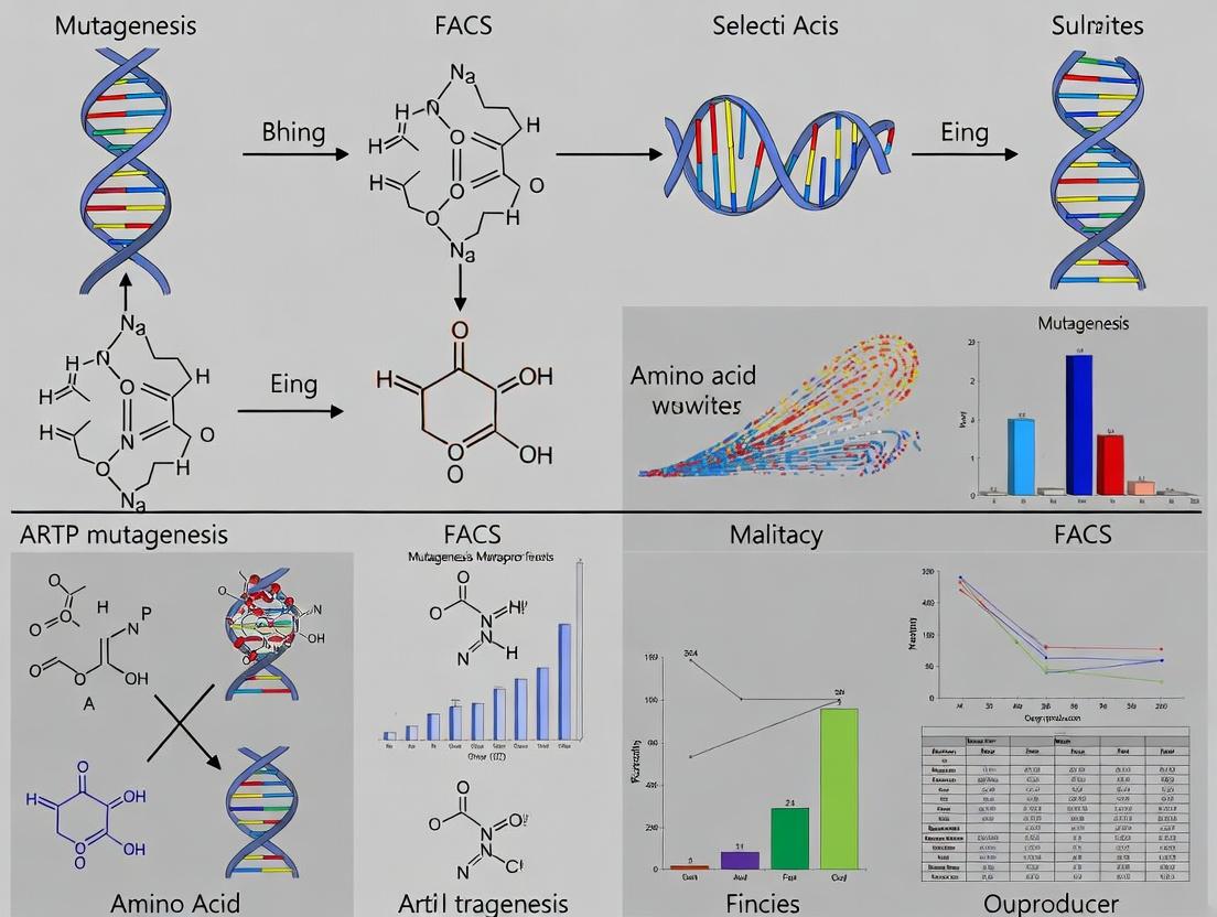
This article provides a comprehensive guide to the integrated workflow of Atmospheric and Room Temperature Plasma (ARTP) mutagenesis and Fluorescence-Activated Cell Sorting (FACS) for the high-throughput selection of microbial strains...
ARTP Mutagenesis and FACS Screening: A High-Throughput Pipeline for Engineering Amino Acid Overproducers
Abstract
This article provides a comprehensive guide to the integrated workflow of Atmospheric and Room Temperature Plasma (ARTP) mutagenesis and Fluorescence-Activated Cell Sorting (FACS) for the high-throughput selection of microbial strains with enhanced amino acid production. Targeting researchers and bioprocess engineers, it covers foundational principles, detailed methodological protocols, common troubleshooting strategies, and validation techniques. The content explores how this combinatorial approach accelerates strain development for industrial fermentation, therapeutic protein manufacturing, and metabolic engineering by efficiently generating and isolating mutants with deregulated biosynthetic pathways. Practical insights into optimizing mutagenesis conditions, designing biosensors for FACS, and benchmarking against alternative methods are included to enable robust implementation in laboratory settings.
Understanding ARTP Mutagenesis and FACS: Core Principles for Strain Improvement
The Need for High-Throughput Strain Engineering in Amino Acid Production
Application Notes: ARTP Mutagenesis and FACS Screening for L-Lysine Overproducers
Thesis Context: This protocol is part of a thesis investigating the synergy of Atmospheric and Room-Temperature Plasma (ARTP) mutagenesis and Fluorescence-Activated Cell Sorting (FACS) to rapidly generate and select microbial strains with enhanced amino acid production, specifically focusing on L-Lysine in Corynebacterium glutamicum.
Rationale: Traditional strain development is slow and low-throughput. This integrated approach accelerates the creation of genetic diversity and enables the screening of tens of thousands of cells to identify rare, high-yielding mutants.
Key Quantitative Data Summary:
Table 1: Typical Mutagenesis and Screening Parameters for C. glutamicum
| Parameter | ARTP Mutagenesis | FACS Screening & Validation |
|---|---|---|
| Mutation Rate Target | 10-30% lethality | N/A |
| Treatment Time | 10-180 seconds | N/A |
| Throughput (Cells/Screen) | ~10^7 total treated | 10^4 - 10^6 cells sorted per hour |
| Mutant Library Size | 10^3 - 10^4 survivors | 0.1 - 1% of population gated |
| Primary Screen Signal | N/A | Fluorescence intensity (A.U.) |
| Yield Improvement (Top Hits) | N/A | 15-45% over parent strain |
| Validation Method | N/A | Shake-flask fermentation (72h) |
Table 2: Example Reagent Solutions for Biosensor-Based FACS
| Reagent/Strain Component | Function in Protocol |
|---|---|
| ARTP Mutagenesis System | Generates reactive plasma species (OH, NO, O) that cause diverse DNA damage/lesions, leading to random mutations. |
| L-Lysine Riboswitch-GFP Biosensor Plasmid | Encodes a GFP reporter under control of a lysine-responsive riboswitch. Intracellular lysine concentration correlates inversely with fluorescence. |
| 96-Well Deep-Well Plates | For high-throughput cultivation of sorted single cells. |
| M9 Minimal Medium + 4% Glucose | Defined medium for selective growth and lysine production during micro-culture validation. |
| O-Phthaldialdehyde (OPA) Derivatization Kit | For high-throughput fluorometric quantitation of L-Lysine in microplate supernatants. |
| Propidium Iodide (PI) Stain | Viability dye for FACS to exclude dead cells from sorting. |
Detailed Protocols
Protocol 1: ARTP Mutagenesis ofC. glutamicum
Objective: Generate a random mutant library with high genetic diversity.
Materials:
- ARTP mutagenesis system
- Wild-type C. glutamicum (ATCC 13032)
- Brain Heart Infusion (BHI) broth and agar plates
- Sterile physiological saline (0.85% NaCl)
- Glass beads or sterile inoculation loop
Method:
- Culture Preparation: Grow the parent strain in 5 mL BHI broth overnight at 30°C, 200 rpm.
- Cell Harvest: Wash cells twice by centrifugation (5000 x g, 5 min) and resuspend in sterile saline to an OD600 of ~1.0.
- Sample Loading: Apply 10 µL of cell suspension onto a sterile, disposable carrier slide. Air-dry in a laminar flow hood for 2-3 minutes.
- Mutagenesis: Place the carrier in the ARTP sample chamber. Set helium gas flow rate to 10 SLM and power to 100 W. Treat sample for a pre-optimized time (e.g., 60 seconds) to achieve ~20% lethality.
- Cell Recovery: Immediately elute treated cells into 1 mL of sterile saline. Perform serial dilution and plate on BHI agar to determine survival rate.
- Library Creation: Dilute the recovered cell suspension and spread on BHI plates to obtain ~500-1000 individual colonies. Incubate at 30°C for 48h. Pick and array colonies into 96-well plates containing BHI to create the mutant master library.
Protocol 2: FACS Screening with a Lysine Biosensor
Objective: Rapidly isolate low-fluorescence mutants indicative of high intracellular lysine.
Materials:
- Mutant library from Protocol 1
- C. glutamicum electrocompetent cells
- Lysine-responsive riboswitch-GFP biosensor plasmid
- FACS sorter (e.g., BD FACSAria III)
- FACS tubes
- LBHIS medium (LB with brain heart infusion and sorbitol)
- Kanamycin (for plasmid maintenance)
- Isopropyl β-D-1-thiogalactopyranoside (IPTG, for biosensor induction)
Method:
- Biosensor Transformation: Electroporate the biosensor plasmid into the pooled mutant library. Select transformations on BHI plates with kanamycin (25 µg/mL). Confirm biosensor response by checking fluorescence reduction with exogenous lysine.
- Induction and Staining: Grow transformed library in 96-well deep plates with BHI+kanamycin for 24h. Subculture into M9 minimal medium + 2% glucose + kanamycin + 0.5 mM IPTG. Grow for 16h to induce biosensor and deplete internal lysine pools. Add propidium iodide (1 µg/mL final) 30 min before sorting to stain dead cells.
- FACS Gating and Sorting:
- Create a dot plot of FSC-A vs SSC-A to gate on the main microbial population.
- Create a PI (e.g., 610/20 filter) vs GFP (530/30 filter) dot plot.
- Gate on PI-negative (live) cells.
- Within live cells, draw a sort gate to collect the bottom 0.5-1% of the population with the LOWEST GFP fluorescence intensity (see Diagram 1).
- Sorting: Sort the low-fluorescence cells directly into a 96-well plate containing 200 µL of recovery medium (BHI) per well. Incubate at 30°C for 48h.
- Validation: Re-screen the sorted populations for stable low fluorescence. Inoculate top candidates from the 96-well plate into 1 mL M9+4% glucose medium in a 96-deep well plate. Ferment for 72h at 30°C, 1000 rpm. Quantify lysine in supernatant using an OPA assay. Scale up top producers in shake flasks for confirmation.
Diagrams
Diagram 1 Title: High-Throughput Strain Engineering Workflow
Diagram 2 Title: FACS Gating Strategy for High Lysine Selection
1. Introduction and Thesis Context This Application Note details Atmospheric and Room Temperature Plasma (ARTP) mutagenesis, a pivotal physical mutagenesis technique for generating microbial genetic diversity. The content is framed within a research thesis focused on developing a high-throughput screening pipeline that integrates ARTP mutagenesis with Fluorescence-Activated Cell Sorting (FACS) to isolate high-titer amino acid overproducing strains. This combinatorial approach addresses the critical bottleneck in microbial strain engineering by coupling broad, random mutagenesis with efficient, targeted screening.
2. Mechanism of ARTP Mutagenesis ARTP utilizes a radio-frequency atmospheric-pressure glow discharge plasma jet to generate a mixture of active particles (e.g., reactive oxygen and nitrogen species, charged particles, UV photons). These agents collectively induce diverse DNA damage in treated cells, including:
- Base damage/modification: Oxidation and nitration of nucleotide bases.
- Single-strand breaks (SSBs) and double-strand breaks (DSBs): Caused by energetic particle bombardment.
- DNA-protein crosslinks.
The cell's error-prone repair mechanisms, such as SOS response in bacteria, introduce mutations during the repair process, leading to a library of genetic variants.
Diagram: Mechanism of ARTP-Induced Mutagenesis
3. Advantages and Comparative Analysis ARTP offers distinct benefits over traditional chemical and physical mutagens.
Table 1: Comparison of Common Mutagenesis Methods
| Feature | ARTP Mutagenesis | UV Mutagenesis | Chemical (EMS/NTG) |
|---|---|---|---|
| Mutation Rate | High (typically 1-30%) | Moderate | High |
| Lethality | Controllable, moderate | High, difficult to control | High, toxic residue risk |
| Operation | Simple, rapid (seconds-minutes) | Simple | Complex, hazardous |
| Safety | High (no toxic chemicals) | High (radiation safety) | Low (carcinogens) |
| Penetration | Good for cell clusters | Poor (surface) | Good |
| Genetic Diversity | Broad, diverse mutation types | Primarily pyrimidine dimers | Primarily point mutations |
| Equipment Cost | Moderate | Low | Very Low |
4. Key Protocols
Protocol 4.1: ARTP Mutagenesis of Amino Acid-Producing Bacteria (e.g., Corynebacterium glutamicum)
A. Materials & Pre-treatment
- Biological Material: Mid-log phase culture of target strain.
- ARTP Instrument: ARTP mutagenesis system (e.g., ARTP-IIS).
- Carrier Plate: Sterile metal carrier plate.
- Solution: Physiological saline (0.85% NaCl) or phosphate buffer.
- Dilution & Plating Media: Appropriate rich and selective agar media.
B. Procedure
- Sample Preparation: Harvest cells by centrifugation. Wash twice and resuspend in saline to a density of ~10⁸ CFU/mL. Pipette 10 µL of suspension onto the center of the sterile carrier plate.
- Instrument Setup: Power on ARTP system. Set helium gas flow rate to 10 slm (standard liters per minute). Set the distance between plasma jet nozzle and sample droplet to 2 mm.
- Mutagenesis Treatment: Start plasma discharge. Treat sample for 10-120 seconds (exact time requires lethality curve optimization). Perform each exposure time in triplicate.
- Post-treatment Recovery: Immediately after treatment, wash the treated cells from the carrier plate into 1 mL of recovery broth. Incubate in the dark at optimal growth temperature with shaking for 2-4 hours to allow expression of mutated phenotypes.
- Lethality Curve Determination: Serially dilute recovered cells and plate on non-selective agar. Count colonies after 24-48 hours. Calculate survival rate = (CFU treated / CFU untreated) * 100%. Plot survival vs. treatment time. For subsequent screening, a treatment time yielding 80-90% lethality is typically optimal.
Protocol 4.2: Integration with FACS for Amino Acid Overproducer Screening
A. Principle: A biosensor or fluorescent reporter system responsive to intracellular amino acid concentration is required. For example, use a transcription factor-based biosensor where target amino acid binding activates GFP expression.
B. Workflow:
Diagram: Integrated ARTP-FACS Screening Pipeline
C. Detailed FACS Protocol:
- Library Preparation: Subject the recovered ARTP mutant pool to cultivation in a defined medium under conditions that couple growth to amino acid production.
- Sample Loading: Dilute or concentrate cells to an event rate of ~10,000 events/second in FACS buffer. Use a strain without the biosensor as a negative control.
- Gating & Sorting: On the flow cytometer, gate on forward/side scatter to exclude debris and aggregates. Set a sorting gate on the far right tail (>99th percentile) of the GFP fluorescence histogram derived from the negative control. Sort the brightest cells directly into 96-well plates containing growth medium.
- Post-Sort Processing: Incubate sorted plates. Screen each well for production titer using a rapid assay (e.g., colorimetric). Validate top performers in shake flasks with analytical quantification (HPLC).
5. The Scientist's Toolkit: Essential Research Reagents & Materials
Table 2: Key Research Reagent Solutions for ARTP-FACS Workflow
| Item | Function in Experiment | Key Consideration |
|---|---|---|
| ARTP Mutagenesis System | Generates plasma for inducing random DNA damage. | Ensure compatibility with anaerobic workstations if needed. Calibrate power and distance. |
| Helium/Nitrogen Gas Supply | Working gas for stable plasma generation. | High purity (>99.99%) required for consistent results. |
| Biosensor Plasmid/Strain | Reports intracellular metabolite level via fluorescence. | Dynamic range, specificity, and lack of metabolic burden are critical. |
| FACS Buffer (PBS + EDTA) | Maintains cell viability and prevents clumping during sorting. | Must be isotonic, filter-sterilized, and may require addition of a carbon source. |
| Fluorescent Protein (e.g., GFP) | The detectable signal for FACS enrichment. | Choose variant with excitation/emission spectra matching your cytometer's lasers/filters. |
| Selective/Screening Media | For outgrowth of sorted cells and preliminary titer assessment. | Formulation should minimize background fluorescence and support product secretion. |
| DNA Repair Inhibitors (Optional) | Enhance mutation frequency by compromising repair fidelity (e.g., caffeine). | Can increase lethality; concentration requires optimization. |
| Analytical Standard (Amino Acid) | For HPLC/LC-MS quantification of final titer. | Use high-purity, isotopically labeled standards for absolute quantification. |
6. Conclusion ARTP mutagenesis is a highly effective tool for generating vast genetic diversity with operational safety and efficiency. When strategically combined with a biosensor-driven FACS screening platform, it forms a powerful closed-loop system for the rapid directed evolution of industrial microbial strains, such as amino acid overproducers, significantly accelerating the strain development timeline.
This application note details the integration of Atmospheric and Room Temperature Plasma (ARTP) mutagenesis with Fluorescence-Activated Cell Sorting (FACS) for high-throughput screening of microbial libraries to select amino acid overproducers. Within the context of a broader thesis, this synergistic approach addresses a key bottleneck in metabolic engineering: rapidly isolating rare, high-performing mutants from vast, diverse libraries. ARTP provides an efficient physical mutagen to generate genetic diversity with low cell toxicity and high mutation rates. Subsequent phenotype-based screening using FACS enables the quantitative, high-speed isolation of cells based on fluorescent biosensor signals linked to intracellular amino acid concentrations, bypassing the limitations of slow, plate-based assays.
Key Protocols
Protocol 1: ARTP Mutagenesis for Microbial Library Creation
- Objective: To generate a diverse mutant library of a microbial strain (e.g., Corynebacterium glutamicum, Escherichia coli) for amino acid overproduction.
- Materials: ARTP mutagenesis system, microbial strain, appropriate liquid growth medium, sterile saline (0.85% NaCl), glass slides, colony counting equipment.
- Detailed Procedure:
- Culture Preparation: Grow the target strain to mid-exponential phase. Harvest cells by centrifugation and wash twice with sterile saline. Adjust cell concentration to ~10⁸ cells/mL.
- Mutagenesis Setup: Place 10 µL of cell suspension on a sterile carrier slide. Insert the slide into the ARTP reactor sample plate.
- Plasma Treatment: Set the plasma jet power to 100-120 W and the helium gas flow rate to 10-12 slm. Treat the sample for a duration determined by a prior kill curve experiment (typically 10-120 seconds). A control (0 seconds) must be included.
- Post-Treatment Recovery: Wash the treated cells from the slide into 1 mL of recovery medium. Incubate with shaking for 2-6 hours for phenotypic expression and recovery.
- Library Preparation: Plate appropriate dilutions to determine survival rate. Use the recovered culture to inoculate main cultures for screening or for cryopreservation of the mutant library.
Protocol 2: FACS Screening Using Fluorescent Biosensors
- Objective: To sort high-performing amino acid-overproducing mutants from an ARTP-generated library using a genetically encoded fluorescent biosensor.
- Materials: FACS sorter (e.g., BD FACSAria, Beckman Coulter MoFlo), microbial library expressing a metabolite-responsive biosensor (e.g., transcription factor-based FRET sensor or single-fluorophore sensor), appropriate growth medium, sterile collection tubes with recovery medium.
- Detailed Procedure:
- Sensor-Strain Preparation: Ensure the ARTP-mutagenized library constitutively expresses a biosensor where fluorescence intensity (e.g., GFP) correlates with intracellular target amino acid concentration.
- Culture & Induction: Grow the mutant library under conditions that promote amino acid production (e.g., nitrogen limitation for lysine). Do not induce sensor expression if it is constitutive.
- Sample Preparation: Harvest cells at mid-exponential phase. Wash and resuspend in FACS buffer (e.g., PBS with minimal glucose). Pass suspension through a cell strainer to remove aggregates.
- FACS Gating & Sorting:
- Use a control strain (low producer) to set the baseline fluorescence.
- Create a scatter gate (FSC vs. SSC) to select single, healthy cells.
- Apply a fluorescence gate to collect the top 0.1%-1% of cells with the highest fluorescence signal.
- Sort these cells in "Enrichment Mode" (first round) or "Single-Cell Mode" (final round) into sterile tubes containing rich recovery medium.
- Recovery & Validation: Incubate sorted cells, then plate on solid medium to obtain single colonies. These are subjected to shake-flask fermentation for HPLC validation of amino acid titer.
Data Presentation
Table 1: Representative Data from ARTP-FACS Screening for L-Lysine Overproducers in C. glutamicum
| Strain / Library Population | Survival Rate Post-ARTP (%) | FACS Fluorescence Gate (Top %) | Sorting Yield (Cells Recovered) | Hit Rate (%)* | Validated L-Lysine Titer (g/L) | Fold Increase vs. WT |
|---|---|---|---|---|---|---|
| Wild-Type (WT) Control | N/A | Baseline | N/A | N/A | 2.1 ± 0.3 | 1.0 |
| ARTP Library (Bulk) | ~25 | N/A (Pre-sort) | N/A | <0.01 | N/D | N/D |
| 1st Sort Enriched Pool | N/A | 1.0 | 5 x 10⁵ | ~1.5 | 3.0 - 4.5 | 1.4 - 2.1 |
| 2nd Sort Single-Cell Clones | N/A | 0.2 | 200 (colonies) | ~85 | 5.8 ± 0.4 (Best Clone) | 2.8 ± 0.2 |
N/A: Not Applicable, N/D: Not Determined. *Hit Rate: Percentage of sorted colonies producing >50% more lysine than WT.
Table 2: Research Reagent Solutions & Essential Materials
| Item Name | Function/Application | Example/Supplier |
|---|---|---|
| ARTP Mutagenesis System | Generates diverse mutant libraries via physical mutagenesis. | ARTP-I/II/III Series (Wuxi Tmaxtree Biotechnology) |
| Fluorescent Biosensor Plasmid | Reports intracellular metabolite concentration as fluorescence. | pSenLys (for lysine) or similar TF-based GFP constructs. |
| FACS Buffer (PBS + 0.1% Glucose) | Maintains cell viability and osmotic balance during sorting. | Prepared in-house or sterile physiological buffer. |
| Cell Strainer (35-70 µm) | Removes cell clumps to prevent FACS nozzle clogging. | Falcon Cell Strainers (Corning). |
| Recovery Medium | Rich, non-selective medium for post-sort cell growth. | Typically BHI or 2xYT for bacteria. |
| HPLC System with UV/FLD | Validates amino acid titers in culture supernatants. | Agilent, Waters, or Shimadzu systems with OPA derivatization. |
Visualizations
Title: ARTP-FACS Screening Workflow for Amino Acid Overproducers
Title: FACS Gating Strategy Using a Fluorescent Biosensor
1. Introduction This document details the application of genetically encoded biosensors for the high-throughput selection of microbial amino acid overproducers. Within the context of a thesis focused on combining ARTP (Atmospheric and Room-Temperature Plasma) mutagenesis with Fluorescence-Activated Cell Sorting (FACS), these biosensors serve as the critical link, converting intracellular metabolite concentration into a quantifiable fluorescent signal. This enables the screening of vast mutant libraries generated by ARTP.
2. Biosensor Design Principles Amino acid biosensors are typically constructed as transcription factor (TF)-based reporter systems. The core components are:
- Sensing Element: A transcription factor (e.g., E. coli’s TrpR for tryptophan, LysG for lysine/arginine) that allosterically binds the target amino acid.
- Reporting Element: A fluorescent protein gene (e.g., sfGFP, mCherry) placed under the control of a promoter regulated by the TF.
- Logic: In a repressor-based system (e.g., TrpR), amino acid binding inactivates the repressor, allowing transcription of the reporter gene. Increased intracellular amino acid concentration thus correlates directly with increased fluorescence.
3. Quantitative Data Summary
Table 1: Common Transcription Factor-Based Biosensors for Amino Acids
| Target Amino Acid | Transcription Factor | Native Organism | Regulatory Logic | Dynamic Range (Fold Induction) | Reported EC₅₀ / KD |
|---|---|---|---|---|---|
| Tryptophan | TrpR | E. coli | Repression | 50-100 | ~5 µM |
| Lysine | LysG | C. glutamicum | Activation | 10-25 | ~1.5 mM |
| Arginine | ArgP | E. coli | Activation | 15-40 | ~100 µM |
| Leucine/Isoleucine/Valine | Lrp | E. coli | Dual (Act./Rep.) | 20-50 (varies by promoter) | ~10 µM (for Leu) |
Table 2: Performance Metrics in a Model Selection Workflow (E. coli)
| Experiment Phase | Typical Library Size | FACS Gate | Enrichment Factor (Over Wild-Type) | Validation Hit Rate (%) |
|---|---|---|---|---|
| Pre-Sort | 10⁹ - 10¹⁰ | N/A | 1 | <0.001 |
| FACS (Top 0.5%) | 5 x 10⁶ | Top FL1 | 200-500 | 15-40 |
| Re-sort / Re-screen | 10⁵ | Top FL1 | >1000 | 60-80 |
4. Detailed Protocols
Protocol 4.1: Construction of a Trp Biosensor Plasmid Objective: Clone the trpR gene and trp promoter (Ptrp) upstream of sfGFP into a medium-copy vector. Materials: pUC19 backbone, genomic DNA from E. coli MG1655, Phusion DNA polymerase, T4 DNA ligase. Procedure:
- Amplify the trpR gene and its native operator/promoter region (Ptrp) using primers that add flanking EcoRI and BamHI sites.
- Digest the pUC19 vector and the PCR product with EcoRI and BamHI. Gel-purify fragments.
- Ligate the Ptrp-trpR fragment into the vector. Transform into competent E. coli. Screen colonies for correct insertion (colony PCR).
- Amplify the sfGFP gene. Clone it downstream of Ptrp using BamHI and HindIII sites, ensuring no transcriptional terminators intervene.
- Sequence the final construct (pBiosensor-Trp) to verify integrity.
Protocol 4.2: Integration of Biosensor into Production Host & Mutant Screening Objective: Generate and screen an ARTP-mutagenized library using FACS. Materials: Production strain (e.g., C. glutamicum ATCC 13032), ARTP mutagenesis system, FACS sorter. Procedure:
- Strain Engineering: Stably integrate the biosensor construct (pBiosensor-Trp) into the chromosome of the production host using site-specific recombination.
- ARTP Mutagenesis: Harvest mid-exponential phase cells, wash, and resuspend in saline. Expose 10 µL of cell suspension on an ARTP sample plate to plasma treatment (e.g., 60-120 seconds). Optimize exposure for ~90% lethality.
- Recovery & Outgrowth: Transfer treated cells to rich recovery medium. Incubate for 4-6 hours. Subsequently, transfer to minimal medium with limited precursor substrate to favor overproducer growth. Outgrow for 24-48 hours.
- FACS Sorting: Dilute cells to ~10⁷ cells/mL in PBS or minimal medium. Sort using a 488 nm laser with a 530/30 nm bandpass filter (for sfGFP). Gate on the top 0.1-1% of fluorescent cells. Collect 10⁶ - 10⁷ cells into recovery medium.
- Validation: Plate sorted cells on solid medium. Pick isolated colonies for shake-flask fermentation. Quantify amino acid titer via HPLC and correlate with fluorescence.
5. Visualizations
Title: Workflow for ARTP-FACS Screening Using Biosensors
Title: Biosensor Activation Pathway
6. The Scientist's Toolkit: Research Reagent Solutions
| Item / Reagent | Function in Biosensor/FACS Workflow | Key Consideration |
|---|---|---|
| ARTP Mutagenesis System | Generates random mutations across the microbial genome via plasma-induced DNA damage. | Critical to calibrate exposure time for optimal mutation rate (~90% lethality). |
| TF-Based Biosensor Plasmid | Encodes the genetic circuit that converts metabolite concentration to fluorescence. | Must be stable (integrated) and have a dynamic range suited to expected overproduction levels. |
| Fluorescent Protein (sfGFP) | The quantitative reporter signal. Its maturation time and brightness are key. | sfGFP is preferred for fast maturation; mCherry allows dual-reporter strategies. |
| FACS Sorter | Physically isolates single cells with the highest fluorescence intensity. | Requires optimization of sheath pressure, nozzle size, and sorting gates for viability. |
| Flow Cytometry Buffer | Suspends cells during analysis without affecting fluorescence or viability. | Typically PBS or minimal medium, may require addition of energy source (e.g., glucose). |
| Amino Acid HPLC Kit | Validates the titer of the target amino acid in culture supernatants post-sort. | Necessary for confirming the correlation between fluorescence and production phenotype. |
| Chromosomal Integration Kit | For stable genomic insertion of the biosensor, avoiding plasmid instability. | Homologous recombination or transposase-based systems (e.g., Tn7) are commonly used. |
Within the ongoing thesis research on ARTP (Atmospheric and Room Temperature Plasma) mutagenesis combined with FACS (Fluorescence-Activated Cell Sorting) for amino acid overproducer selection, the derived microbial strains have profound applications. This work bridges foundational strain development with critical industrial and pharmaceutical bioprocessing. The overproduction of amino acids like L-tryptophan, L-tyrosine, and L-lysine serves as a cornerstone for both bulk fermentation and the synthesis of high-value drug precursors.
Application Notes
Industrial Fermentation for Amino Acid Production
Optimized overproducer strains developed via ARTP/FACS are deployed in large-scale fed-batch fermenters. Key performance metrics from recent scale-up trials are summarized below.
Table 1: Performance Metrics of Amino Acid Overproducers in Industrial Fermentation
| Amino Acid | Host Strain | Final Titer (g/L) | Yield (g/g Glucose) | Productivity (g/L/h) | Fermentation Scale (L) |
|---|---|---|---|---|---|
| L-Lysine HCl | C. glutamicum AHP-7 | 185 | 0.55 | 2.57 | 50,000 |
| L-Tryptophan | E. coli TRP-12 | 68 | 0.23 | 0.94 | 30,000 |
| L-Tyrosine | E. coli TYR-9 | 55 | 0.19 | 0.76 | 15,000 |
Precursor Synthesis for Drug Development
Specific amino acid overproducers serve as chassis for the synthesis of complex pharmaceutical precursors. For instance, L-tyrosine overproducers are engineered to express additional plant-derived enzymes for the biosynthesis of L-DOPA, a critical drug for Parkinson's disease. Similarly, tryptophan overproducers are diverted into pathways for indole alkaloid precursor synthesis.
Table 2: Drug Precursor Synthesis from Amino Acid Overproducers
| Target Precursor | Parent Amino Acid | Engineered Pathway | Key Heterologous Enzyme(s) | Precursor Titer (mg/L) |
|---|---|---|---|---|
| L-DOPA | L-Tyrosine | Tyrosine Hydroxylation | Tyrosine hydroxylase (AtTyrH) | 1,450 |
| 4-Hydroxy-L-phenylglycine | L-Tyrosine | Hydroxylation & Transamination | p-hydroxymandelate synthase (HmaS) | 890 |
| Halogenated Tryptophan Derivatives | L-Tryptophan | Tryptophan Halogenation | Tryptophan 6-halogenase (SttH) | 620 (6-Cl-Trp) |
Detailed Protocols
Protocol 1: ARTP Mutagenesis of Amino Acid-Producing Bacteria
Objective: To generate genetic diversity in a bacterial population for enhanced amino acid production.
Materials:
- ARTP mutagenesis system (Wuxi Tmaxtree Biotechnology)
- Late-log phase bacterial culture (OD600 ~0.8)
- Sterile physiological saline (0.85% NaCl)
- Appropriate agar plates for recovery and selection.
Procedure:
- Harvest 10 mL of late-log phase cells by centrifugation (5,000 x g, 4°C, 5 min).
- Wash cell pellet twice with 10 mL sterile physiological saline and resuspend in saline to a final concentration of ~10⁸ cells/mL.
- Pipette 10 µL of cell suspension onto a sterile metal carrier slide. Air-dry in a laminar flow hood for 3-5 minutes.
- Insert the slide into the ARTP reactor chamber. Treat the cells with helium plasma (power: 100 W, gas flow rate: 10 slm, treatment distance: 2 mm). Critical: Perform a kill curve analysis first. Typical treatment times range from 10-120 seconds, aiming for a survival rate of 10-30%.
- Post-treatment, immediately elute the cells from the slide with 1 mL of saline. Perform serial dilutions.
- Plate appropriate dilutions on non-selective recovery agar. Incubate for 24-48 hours.
- Use colonies from the recovery plates to inoculate a master plate for subsequent screening via FACS.
Protocol 2: FACS Screening for Amino Acid Overproduction Using Biosensors
Objective: To high-throughput screen the ARTP-mutagenized library for clones with enhanced amino acid synthesis.
Materials:
- Fluorescence-activated cell sorter (e.g., BD FACSAria III)
- Bacterial library from Protocol 1.
- Growth medium supplemented with a fluorescence-inducing agent (e.g., anhydrotetracycline for biosensor induction).
- Amino acid-responsive transcriptional biosensor plasmid (e.g., pSenLys for lysine).
Procedure:
- Transform the amino acid-specific transcriptional biosensor plasmid into the ARTP-mutagenized library pool. The biosensor links intracellular amino acid concentration to GFP expression.
- Grow the transformed library in 96-deep well plates for 16-20 hours in selective medium.
- Dilute cultures 1:100 in fresh medium containing the biosensor inducer. Grow to mid-log phase (OD600 ~0.5).
- Dilute cells 1:10 in PBS or sorting buffer. Filter through a 35 µm cell strainer.
- Configure the FACS sorter: Use a 488 nm laser for excitation and a 530/30 nm bandpass filter for GFP detection.
- Set sorting gates based on fluorescence intensity of a wild-type control population. Gate the top 0.1-1% of highly fluorescent cells.
- Sort the positive population into sterile microcentrifuge tubes containing recovery medium.
- Plate sorted cells on selective agar to obtain single colonies. Validate amino acid titer in shake-flask fermentation.
Protocol 3: Fed-Batch Fermentation for L-Lysine Production
Objective: To scale up production from a selected overproducer strain.
Materials:
- Seed culture of C. glutamicum AHP-7.
- Fermentation basal salts medium (glucose, (NH4)2SO4, KH2PO4, MgSO4·7H2O, trace elements, vitamins).
- 50 L Bioreactor with automated pH, dissolved oxygen (DO), and temperature control.
- Antifoam agent, 50% (w/v) glucose feed solution, 25% (v/v) ammonium hydroxide solution.
Procedure:
- Inoculate a 1 L shake flask with a colony and grow for 16 hours at 30°C, 220 rpm.
- Transfer the seed culture to the bioreactor containing 30 L of basal medium. Initial conditions: 30°C, pH 7.0 (controlled with NH4OH), DO maintained at 30% saturation via cascaded agitation and aeration.
- Initiate the glucose feed when the initial batch glucose is depleted (typically after 12-16 hours). Maintain glucose concentration at 5-20 g/L using a pre-programmed exponential feed profile.
- Fermentation runs for ~72 hours. Sample periodically for OD600, residual glucose, and lysine quantification (HPLC).
- Terminate fermentation when the lysine titer plateaus. Cool the broth and harvest.
The Scientist's Toolkit
Table 3: Key Research Reagent Solutions
| Item / Reagent | Function / Application |
|---|---|
| ARTP Mutagenesis System | Delivers helium plasma to induce random DNA damage and mutations in microbial genomes. |
| Fluorescent Biosensor Plasmids (e.g., pSenLys) | Genetically encoded reporters that couple intracellular metabolite concentration to GFP signal for FACS. |
| Defined Fermentation Medium | Provides optimized salts, vitamins, and carbon source for reproducible, high-yield amino acid production. |
| HPLC with UV/FLD Detector | Quantifies amino acid concentrations in fermentation broth and screening samples. |
| Fluorescence-Activated Cell Sorter (FACS) | Enables high-throughput, quantitative screening of millions of cells based on fluorescence intensity. |
| 96-Deep Well Plate System | Allows parallel miniaturized cultivation of mutant libraries prior to FACS analysis. |
| Electroporator & High-Efficiency Competent Cells | For transformation of biosensor plasmids into mutagenized libraries. |
Diagrams
ARTP-FACS Workflow for Overproducer Development
L-DOPA Biosynthesis from L-Tyrosine
Key Applications of Developed Overproducer Strains
Step-by-Step Protocol: Combining ARTP and FACS to Isolate Overproducers
This document details the critical pre-mutagenesis steps of strain selection and cultivation within a broader thesis research framework aiming to develop high-throughput microbial cell factories. The core methodology integrates Atmospheric and Room Temperature Plasma (ARTP) mutagenesis with Fluorescence-Activated Cell Sorting (FACS) for the efficient screening of amino acid overproducers. The fitness and genetic background of the starting strain, coupled with precise pre-cultivation conditions, are paramount to the success of the subsequent mutagenesis and high-throughput screening pipeline.
Strain Selection: Criteria and Rationale
Selecting an appropriate parental strain is the first decisive step. The criteria must align with the end goal of amino acid overproduction.
Table 1: Key Criteria for Parental Strain Selection
| Criterion | Explanation & Rationale | Quantitative Target/Example |
|---|---|---|
| Genomic Stability | Low spontaneous mutation rate to ensure ARTP-induced variants are primary contributors. | Mutation rate < 1 x 10⁻⁹ per base per generation. |
| Genetic Tractability | Ease of genetic manipulation for later pathway engineering or reporter gene insertion. | Availability of established transformation protocols (e.g., electrocompetent cells). |
| Robust Growth | Fast, reproducible growth in defined media to ensure consistent pre-cultivation for ARTP. | Doubling time < 1 hour in log phase (for bacteria). |
| Amino Acid Pathway | Possession of a native, strong promoter for the target amino acid's biosynthetic pathway. | Known genomic sequence of operons (e.g., ilv for branched-chain, lys for lysine). |
| Safety & Containment | Generally Recognized As Safe (GRAS) status or BSL-1 classification for lab safety. | Strains: Corynebacterium glutamicum, Bacillus subtilis, Escherichia coli K-12. |
| Previous Yield Baseline | Documented, low-level production of the target amino acid to provide a baseline for improvement. | Measurable titer in flask culture (e.g., 0.5 - 2.0 g/L L-lysine). |
Cultivation Protocols for Pre-Mutagenesis
Standardized cultivation is essential to obtain a homogeneous, physiologically active cell population optimal for ARTP treatment.
Protocol: Preparation of Seed Culture
Objective: To generate an actively growing, homogeneous inoculum.
- Medium: Use a defined, minimal medium (e.g., M9 for E. coli, CGXII for C. glutamicum) with a low, growth-limiting concentration of the target amino acid (e.g., 0.1 g/L) to pre-adapt metabolism.
- Inoculation: Scrape a single colony from a fresh (<48h) agar plate into 5 mL of medium in a 15 mL tube.
- Incubation: Culture at strain-optimal temperature (e.g., 30°C for C. glutamicum, 37°C for E. coli) with shaking at 220 rpm for 6-8 hours (to early log phase, OD600 ≈ 0.3-0.5).
Protocol: Preparation of Main Culture for ARTP
Objective: To scale up culture to the required biomass in a controlled physiological state.
- Dilution: Dilute the seed culture into 50 mL of fresh, pre-warmed minimal medium in a 250 mL baffled flask to an initial OD600 of 0.05.
- Growth Monitoring: Incubate under optimal conditions, monitoring OD600 every 30-60 minutes.
- Harvest Point: Harvest cells at mid-log phase (OD600 ≈ 0.6-0.8). This ensures maximum cell wall permeability and metabolic activity, which correlates with higher mutagenesis efficiency.
- Cell Washing: Centrifuge culture (4,000 x g, 4°C, 10 min). Wash pellet twice with sterile, cold 0.9% (w/v) NaCl solution or phosphate buffer (pH 7.0) to remove medium components that could interfere with plasma treatment.
- Final Suspension: Resuspend the final pellet in the same saline/buffer solution to a standardized cell density. Critical Density:
1.0 x 10^8 to 5.0 x 10^8 cells/mL(approximately OD600 = 0.5-1.0 for most bacteria). Keep suspension on ice until ARTP treatment (within 30 min).
Table 2: Standardized Pre-Mutagenesis Culture Conditions
| Parameter | Condition for E. coli | Condition for C. glutamicum | Purpose |
|---|---|---|---|
| Medium | M9 Minimal + 0.1% Glu | CGXII Minimal + 0.1% Glu | Defined conditions, induces biosynthetic pathways. |
| Temperature | 37°C | 30°C | Optimal for growth rate and physiology. |
| Harvest OD600 | 0.6 ± 0.05 | 0.7 ± 0.05 | Mid-log phase cells are most susceptible to mutagenesis. |
| Wash Buffer | 0.1M PBS (pH 7.2) | 0.9% NaCl | Removes ions, standardizes ionic environment. |
| Final Density | 5.0 x 10⁸ cells/mL | 3.0 x 10⁸ cells/mL | Optimal monolayer formation on ARTP carrier slide. |
Visualizing the Integrated Workflow
Title: Integrated ARTP-FACS Workflow for Amino Acid Producer Development
The Scientist's Toolkit: Research Reagent Solutions
Table 3: Essential Materials and Reagents for Pre-Mutagenesis Preparation
| Item | Function / Purpose | Example Product/Catalog |
|---|---|---|
| Defined Minimal Media | Provides controlled, reproducible growth conditions without complex additives, forcing reliance on native amino acid biosynthesis. | M9 Broth (Sigma-Aldrich, M6030), CGXII Salts. |
| Sterile Saline (0.9% NaCl) | Isotonic solution for washing and resuspending cells post-harvest to remove growth medium and standardize samples for ARTP. | Sterile-filtered 0.9% NaCl solution (Lab-prepared). |
| Phosphate Buffered Saline (PBS) | Maintains pH and osmotic balance during cell washing, crucial for maintaining cell viability pre-mutagenesis. | 1X PBS, pH 7.4 (Gibco, 10010023). |
| Baffled Erlenmeyer Flasks | Enhances oxygen transfer during pre-cultivation, ensuring aerobic growth and preventing metabolic shifts to fermentation. | 250 mL Baffled Flask (Corning, 4450-0250). |
| Spectrophotometer & Cuvettes | For precise optical density (OD600) measurements to monitor growth and standardize cell density for ARTP treatment. | NanoDrop One⁺ (Thermo Fisher) or equivalent. |
| Refrigerated Benchtop Centrifuge | For pelleting microbial cells gently and quickly at 4°C to maintain viability and halt metabolic activity at harvest. | Eppendorf 5430 R with rotor for 50 mL tubes. |
| ARTP Mutagenesis System | The mutagenesis instrument generating the reactive plasma species (ROS, RNS, UV) that cause DNA damage and mutations. | ARTP-IIS or ARTP-M Biological Mutagenesis Instrument. |
| Sterile Carrier Slides (Quartz) | Platform on which the cell suspension is placed as a thin film for uniform exposure to the plasma jet. | 15 mm diameter quartz slides, sterilized. |
Atmospheric and Room Temperature Plasma (ARTP) mutagenesis is a powerful, non-GM physical technique for microbial breeding, inducing DNA damage via reactive species. Within a thesis integrating ARTP with Fluorescence-Activated Cell Sorting (FACS) for amino acid overproducer selection, precise optimization of ARTP parameters (exposure time, helium flow rate) is critical to achieve a high mutation rate with a suitable survival rate, generating a diverse mutant library for downstream high-throughput screening. This protocol details the systematic optimization process.
ARTP Mutagenesis Mechanism
The ARTP system generates a plasma jet at room temperature using radio-frequency power and a helium gas flow. The plasma contains chemically active species (e.g., ·OH, ·O, excited He) that cause diverse DNA lesions (base damage, single/double-strand breaks). Optimal parameters balance DNA damage intensity with cell repair capacity, maximizing genetic diversity while maintaining sufficient viable cells for FACS screening.
Parameter Optimization Protocol
Materials and Equipment
Research Reagent Solutions:
| Item | Function |
|---|---|
| ARTP Mutation System (e.g., ARTP-I/II/III) | Core equipment generating the helium plasma jet at room temperature. |
| Helium Gas (≥99.999% purity) | Plasma working gas; purity ensures consistent reactive species generation. |
| Microbial Strain (e.g., Corynebacterium glutamicum) | Target microorganism for amino acid overproduction. |
| Phosphate Buffered Saline (PBS, 0.1M, pH 7.0-7.4) | Suspension buffer for cells during treatment to maintain isotonic conditions. |
| Appropriate Solid/Liquid Growth Media | For pre-culture, post-treatment recovery, and survival rate calculation. |
| Sterile Inoculation Loop or Cell Spreaders | For plating and colony counting. |
| Anaerobic Jar/Bag (if required) | For strains requiring specific atmospheres during recovery. |
Pre-treatment Preparation
- Culture Preparation: Inoculate the target strain into liquid medium. Incubate to mid-logarithmic growth phase (OD~600~ ~0.6-0.8). Harvest cells by centrifugation (e.g., 5000 rpm, 5 min).
- Cell Washing & Suspension: Wash cell pellet twice with sterile PBS. Re-suspend to a density of ~10⁸ cells/mL (critical for consistent plasma interaction). Keep on ice.
- Sample Loading: Aliquot 10 µL of cell suspension onto a sterile metal carrier slide. Gently spread to form a thin film. Air-dry in a sterile laminar flow hood for 3-5 minutes.
Experimental Design for Optimization
A two-factor central composite design or full factorial design is recommended. Core test ranges (based on current literature):
- Helium Flow Rate: 8 - 16 standard liters per minute (slm).
- Exposure Time: 10 - 180 seconds.
- Fixed Parameters: RF power input (e.g., 100-120 W), electrode distance (~2 mm), treatment volume (10 µL).
Procedure:
- System Setup: Power on the ARTP instrument and gas supply. Set the RF power to the fixed value. Purge the system with helium at the desired test flow rate for 1 minute to stabilize.
- Plasma Treatment: Place the sample slide under the plasma jet at the fixed distance. Initiate treatment for the predetermined exposure time. Each parameter combination should be performed in triplicate.
- Post-treatment Recovery: Immediately after treatment, wash the cells from the slide with 1 mL of recovery medium (or PBS). Perform serial dilutions (10⁻¹ to 10⁻⁶) in PBS.
- Viability Assay: Plate 100 µL of appropriate dilutions onto solid agar plates. Incubate under optimal growth conditions for 24-48 hours.
- Control: Treat a sample with helium flow but no plasma discharge (0 seconds) as a negative control.
Data Collection & Analysis
- Survival Rate Calculation: Survival Rate (%) = (CFU/mL of treated sample / CFU/mL of control sample) × 100%.
- Target Survival Window: For mutant library construction, aim for a survival rate between 10% and 30%. This typically yields a high mutation frequency (10⁻³ to 10⁻⁵) without excessive cell death.
- Mutation Frequency Validation: Randomly pick 100-200 survivors from plates corresponding to the target survival window. Screen for a desired phenotype (e.g., amino acid analog resistance) or use genetic methods (RAPD, sequencing) to estimate DNA mutation frequency.
Optimized Parameter Integration with FACS Workflow
The optimized ARTP conditions are the first step in the integrated thesis pipeline. Survivors are recovered in bulk and used to inoculate a fermentation culture. Subsequent FACS screening is based on biosensor fluorescence (for intracellular amino acid concentration) or proxy indicators.
Table 1: Representative ARTP Parameter Effects on Microbial Survival Rates
| Strain Type | Helium Flow (slm) | Exposure Time (s) | Avg. Survival Rate (%) | Typical Mutation Frequency | Reference Context |
|---|---|---|---|---|---|
| C. glutamicum | 10 | 30 | 85.2 ± 3.1 | ~10⁻⁵ | Preliminary, low lethality |
| C. glutamicum | 12 | 90 | 22.5 ± 4.7 | ~10⁻⁴ | Optimal Library Range |
| C. glutamicum | 12 | 120 | 8.1 ± 2.3 | ~10⁻³ | High lethality, high diversity |
| E. coli | 10 | 60 | 18.7 ± 3.5 | ~10⁻⁴ | Comparative benchmark |
| S. cerevisiae | 15 | 120 | 15.0 ± 5.0 | ~10⁻⁴ | Eukaryotic example |
Table 2: Key Reagents and Materials for ARTP-FACS Pipeline
| Step | Key Solution/Material | Specification/Function |
|---|---|---|
| ARTP Treatment | Helium Gas | High purity (99.999%) for stable plasma generation. |
| Cell Handling | Phosphate Buffered Saline (PBS) | Ionic strength maintains cell integrity during treatment. |
| Recovery | Rich Medium (e.g., BHI for bacteria, YPD for yeast) | Supports repair of sub-lethally injured cells post-ARTP. |
| FACS Staining | Fluorescent Biosensor (e.g., GFP-based transcription factor sensor) | Reports intracellular metabolite (amino acid) levels. |
| FACS Buffer | Cell Staining Buffer (PBS + 0.5% BSA) | Reduces non-specific binding and cell clumping for sorting. |
| Culture | Defined/Analogue Media | For selective outgrowth of overproducing mutants post-FACS. |
Visualization
ARTP Optimization & FACS Screening Workflow
ARTP Parameter Impact on DNA & Cell Fate
Constructing and Calibrating Fluorescent Biosensors for Target Amino Acids
This protocol details the construction and calibration of genetically encoded fluorescent biosensors for intracellular amino acid quantification. Within the broader thesis on developing microbial strains for amino acid overproduction, these biosensors serve as the critical phenotype-genotype link. Following ARTP (Atmospheric and Room Temperature Plasma) mutagenesis to generate genetic diversity, these sensors enable high-throughput screening via Fluorescence-Activated Cell Sorting (FACS). Isolated high-fluorescence cells correspond to mutants with elevated target amino acid titers, directly linking biosensor output to production phenotype.
Application Notes & Core Principles
Biosensor Design: Modern biosensors for amino acids are typically based on transcription factor-based Forster Resonance Energy Transfer (FRET) sensors or single fluorescent protein (FP) insertion-based sensors. The sensing element is a bacterial periplasmic binding protein (PBP) or a eukaryotic amino acid receptor domain, which undergoes a conformational change upon ligand binding. This change is transduced into a change in fluorescence intensity or FRET ratio. Key Considerations: Dynamic range, specificity (minimal cross-reactivity with analogous amino acids), affinity (Kd should match the expected physiological concentration range), brightness, and response kinetics are critical. Sensors must be expressed in the host production strain (e.g., Corynebacterium glutamicum, Escherichia coli) without disrupting metabolism.
Detailed Experimental Protocols
Protocol 3.1: Molecular Construction of a FRET-Based Biosensor
Objective: Clone a genetically encoded FRET biosensor for L-Lysine into an appropriate expression vector. Materials: See "Research Reagent Solutions" table. Procedure:
- Template Amplification: Using PCR, amplify the coding sequence for a lysine-binding protein (e.g., E. coli LysP or a designed variant) from genomic DNA or a synthetic gene.
- Vector Preparation: Digest the mammalian or bacterial expression vector (e.g., pRSETB, pET-based for bacteria; pcDNA3 for mammalian cells) with appropriate restriction enzymes (e.g., BamHI and EcoRI). Gel-purify the linearized vector.
- FP Fusion:
- Perform overlap extension PCR to fuse the amplified binding protein sequence between the genes for the donor FP (e.g., ECFP, mCerulean3) and the acceptor FP (e.g., Venus, mCitrine).
- Ensure linkers (typically 5-15 aa, e.g., GGGGS repeats) are included between the binding protein and each FP to permit conformational freedom.
- Gibson Assembly/Ligation: Assemble the FP-binding protein-FP fragment into the prepared vector using Gibson Assembly or traditional ligation.
- Transformation & Verification: Transform the assembly into competent E. coli (e.g., DH5α). Screen colonies by colony PCR and verify the final plasmid by Sanger sequencing.
Protocol 3.2:In VitroPurification and Affinity Calibration (Kd Determination)
Objective: Purify the biosensor protein and determine its dissociation constant (Kd) for the target amino acid. Procedure:
- Expression & Purification: Transform the biosensor plasmid into a protein expression host (e.g., BL21(DE3)). Induce expression with IPTG. Lyse cells and purify the His-tagged biosensor via Ni-NTA affinity chromatography.
- Fluorescence Measurement Setup: Prepare a dilution series of the purified biosensor (e.g., 100 nM) in a suitable buffer (e.g., PBS, pH 7.4) in a 96-well plate or cuvette.
- Titration: For a FRET sensor, add increasing concentrations of the target amino acid (e.g., 0, 0.1, 0.5, 1, 5, 10, 50, 100, 500 µM L-Lysine). For an intensity-based sensor, use the relevant excitation/emission wavelengths.
- Data Acquisition: Measure the donor emission, acceptor emission, and calculate the FRET ratio (Acceptor Emission / Donor Emission) at each ligand concentration. Perform triplicate measurements.
- Curve Fitting: Plot the FRET ratio (or normalized fluorescence intensity) against the log of amino acid concentration. Fit the data to a sigmoidal dose-response curve (e.g., using a four-parameter logistic equation in GraphPad Prism or similar) to determine the Kd (ligand concentration at half-maximal response).
Protocol 3.3:In VivoCalibration in Microbial Hosts
Objective: Characterize biosensor performance in the actual production host strain (e.g., C. glutamicum). Procedure:
- Strain Engineering: Introduce the biosensor expression vector into the wild-type or ARTP-mutagenized host strain via electroporation or conjugation.
- Calibration Culture: Grow sensor-expressing cells in minimal medium to mid-exponential phase.
- External Calibration: Aliquot cells and treat with a range of known concentrations of the target amino acid (0-100 mM) in the presence of a membrane permeabilizer (e.g., 0.1% toluene/ethanol mix for E. coli; 0.01% digitonin for C. glutamicum) for 10-15 minutes. This equilibrates intra- and extracellular concentrations.
- Flow Cytometry Analysis: Analyze each sample by flow cytometry, gating on healthy cells. Record the mean fluorescence intensity (for intensity sensors) or ratiometric values (for FRET sensors) for the population.
- Internal Standard Curve: Plot the cellular fluorescence/ratio against the known external amino acid concentration to generate an in vivo standard curve. This curve is used to convert FACS fluorescence data from mutant libraries into estimated intracellular amino acid concentrations.
Data Presentation
Table 1: Example In Vitro Calibration Data for a Lysine FRET Biosensor
| Ligand (Lysine) Concentration (µM) | Mean FRET Ratio (A.U.) | Standard Deviation (n=3) | Normalized Response (%) |
|---|---|---|---|
| 0 | 1.05 | 0.03 | 0 |
| 1 | 1.08 | 0.04 | 4.5 |
| 5 | 1.25 | 0.05 | 25.6 |
| 20 | 1.63 | 0.07 | 74.1 |
| 100 | 1.85 | 0.06 | 100 |
| 500 | 1.86 | 0.05 | 101 |
| Fitted Kd (µM) | 18.5 ± 1.2 | ||
| Dynamic Range (Rmax/Rmin) | 1.77 |
Table 2: Key Research Reagent Solutions
| Item | Function/Explanation | Example Product/Catalog # |
|---|---|---|
| ARTP Mutagenesis System | Generates random genomic mutations in microbial cells to create diverse mutant libraries. | ARTP-M Microbial Mutagenesis System |
| Fluorescent Protein Genes | Donor/Acceptor pairs for FRET (e.g., mCerulean3/mCitrine) or single bright FPs (e.g., sfGFP). | Clontech FP vectors; Addgene plasmids #54529, #54531 |
| Periplasmic Binding Protein (PBP) Domains | The sensing element; confers specificity for the target amino acid. | E. coli LysP (Lysine), GlnH (Glutamine); S. cerevisiae Gap1 (general amino acid). |
| High-Fidelity Polymerase | For error-free amplification of biosensor gene fragments. | Q5 High-Fidelity DNA Polymerase (NEB) |
| Gibson Assembly Master Mix | Enables seamless, single-step assembly of multiple DNA fragments. | Gibson Assembly HiFi Master Mix (NEB) |
| Ni-NTA Agarose Resin | For purification of polyhistidine (His)-tagged biosensor proteins. | Ni-NTA Superflow (QIAGEN) |
| Membrane Permeabilizer | Allows controlled access of external amino acids to the cytosol for in vivo calibration. | Digitonin (Sigma D141) |
| Flow Cytometer with Cell Sorter | For high-throughput analysis and sorting of sensor-expressing cell populations. | BD FACS Aria, Beckman Coulter MoFlo Astrios |
Mandatory Visualizations
Diagram Title: Biosensor-Driven FACS Selection Workflow for Amino Acid Overproducers
Diagram Title: FRET Biosensor Mechanism Upon Amino Acid Binding
Within the research framework of coupling ARTP (Atmospheric and Room Temperature Plasma) mutagenesis with FACS (Fluorescence-Activated Cell Sorting) for the high-throughput selection of microbial amino acid overproducers, the FACS workflow is the critical linchpin. This protocol details the steps to effectively screen vast mutant libraries generated by ARTP, where random mutagenesis creates genetic diversity. The workflow hinges on coupling amino acid overproduction to a fluorescent reporter, enabling the isolation of rare high-producing variants via FACS. Rigorous gating, optimized sorting parameters, and careful post-sort recovery are essential to ensure the isolation of viable, genetically stable overproducers for downstream characterization in drug development and biomanufacturing pathways.
Key Research Reagent Solutions
| Reagent/Material | Function in ARTP-FACS Workflow |
|---|---|
| ARTP Mutagenesis System | Generates random mutations in microbial genomes to create genetic diversity for screening. |
| Fluorescent Biosensor | A reporter system (e.g., transcription factor-based or FRET-based) that changes fluorescence intensity in response to intracellular amino acid concentration. |
| Cell Viability Stain (e.g., PI) | Distinguishes live from dead cells during gating; critical for sorting only viable mutants. |
| Sterile Sheath Fluid | The isotonic, particle-free fluid that hydrodynamically focuses the cell stream in the sorter. |
| High-Recovery Growth Medium | Enriched, osmotically balanced medium used for sample collection and post-sort recovery to minimize stress. |
| Antibiotic/Antifungal (optional) | Added to collection medium to prevent contamination during long sort sessions. |
| 96- or 384-well Plate Pre-filled with Medium | For single-cell deposition and clonal outgrowth post-sort. |
Detailed FACS Protocol for Mutant Screening
Sample Preparation Pre-Sort
- Induction: Induce the fluorescent biosensor reporter in the ARTP-mutagenized cell library according to its specific mechanism (e.g., limit specific amino acid to induce sensor response).
- Washing & Resuspension: Harvest cells by gentle centrifugation. Wash twice and resuspend at a final density of 1-5 x 10^6 cells/mL in sterile PBS or appropriate sorting buffer.
- Filtration: Pass cell suspension through a 35-70 µm cell strainer to remove aggregates that can clog the instrument.
- Viability Staining (Optional but Recommended): Add a viability dye (e.g., Propidium Iodide, 1-5 µg/mL) and incubate for 5-10 minutes on ice. Protect from light.
Instrument Setup & Gating Strategy
- Calibration: Perform daily startup and calibration using standardized beads for fluidics, lasers, and optical alignment.
- Trigger & Threshold: Set the primary trigger parameter to Forward Scatter (FSC) to ignore small debris. Adjust threshold appropriately.
- Gating Hierarchy: Implement the following sequential gating logic visualized in the diagram below.
Diagram Title: Sequential Gating Strategy for ARTP-FACS Mutant Screening
Sorting Parameters Configuration
Critical sorting parameters must be balanced to achieve purity, viability, and efficiency.
| Parameter | Recommended Setting | Purpose & Rationale |
|---|---|---|
| Nozzle Size | 70-100 µm | For microbial cells; balances shear stress (viability) and sorting speed. |
| Sheath Pressure | 20-25 psi (for 70µm) | Lower pressure favors viability. Adjust with nozzle size. |
| Sort Mode | Purity (for single-cell cloning) | Prioritizes purity of the sorted population over yield. |
| Drop Delay | Precisely determined daily using beads | Critical for sort accuracy; misalignment causes failed sorts. |
| Sorting Speed | 200-1000 events/sec* | Kept low to maintain high sort efficiency and viability. |
| Collection Device | 96-well plate with medium | Enables clonal outgrowth directly from sorted single cells. |
| Sorting Enrichment | Top 0.1% - 1% of Sensor+ population | Isolates the extreme tail of the fluorescence distribution. |
Note: Speed depends on cell type, density, and desired recovery.
Post-Sort Recovery Protocol
- Immediate Processing: Post-sort, seal collection plates and transfer to appropriate growth conditions (incubator/shaker) within 30 minutes.
- Outgrowth: Incubate without disturbance for 24-48 hours to allow clonal growth from single cells.
- Re-screening: Use a small aliquot from each well for a confirmatory analytical assay (e.g., microplate fluorescence reader) to identify true positive overproducers before colony expansion.
- Expansion & Validation: Expand positive clones for validation using HPLC or LC-MS to quantitatively measure amino acid titers, ensuring linkage between fluorescence and production phenotype.
| Workflow Phase | Key Metric | Typical Target/Outcome | Impact on Selection |
|---|---|---|---|
| Pre-Sort | Mutant Library Size | >10^7 independent mutants | Ensures sufficient diversity for rare high-producers. |
| Gating | Live Cell Recovery | >80% of total events | Maximizes viable candidates for sorting. |
| Sorting | Sort Efficiency (Purity) | >95% (in Purity mode) | Ensures single-cell cloning fidelity. |
| Sorting | Event Rate | <10,000 events/sec | Maintains sort accuracy and cell viability. |
| Post-Sort | Well Occupancy (Single-cell) | ~0.5 cells/well (for 96-well) | Optimizes for clonality vs. throughput. |
| Post-Sort | Clonal Outgrowth Rate | >70% of sorted wells | Indicates maintenance of cell viability through process. |
| Validation | False Positive Rate | <20% (after re-screen) | Determined by correlation of fluorescence with final product titer. |
Diagram Title: Integrated ARTP Mutagenesis and FACS Screening Workflow
1. Introduction & Context within ARTP-FACS Thesis This protocol details the critical validation step following a high-throughput screening campaign for amino acid overproducers. Within the broader thesis on coupling ARTP Mutagenesis with FACS-based selection, initial hits are identified via fluorescence biosensors or growth-coupled selection in microtiter plates. This document describes the systematic process of transitioning these primary hits from 96-well plates to small-scale shake flask fermentation to confirm production phenotypes under more physiologically relevant conditions, eliminating false positives from plate-based artifacts.
2. Experimental Protocol: Tiered Validation Workflow
2.1. Protocol A: Primary Hit Confirmation in 96-Well Plates Objective: Re-evaluate initial FACS-sorted clones for reproducible production and growth. Methodology:
- Inoculum Preparation: Pick individual colonies from sorted/plated populations into 150 µL of defined minimal medium in a 96-well deep-well plate (2 mL capacity). Culture for 48 hours at 30°C, 850 rpm.
- Micro-cultivation: Transfer 10 µL of pre-culture into 190 µL of fresh production medium in a standard 96-well plate. Incubate for 72 hours at 30°C, 900 rpm in a controlled-climate shaker.
- Analytical Sampling: At 24, 48, and 72 hours, measure:
- OD600: For growth kinetics.
- Fluorescence (if applicable): For biosensor-based hits (Ex/Em as per biosensor).
- Supernatant Analysis: Using a minimum volume assay (e.g., NADH-linked enzymatic assays for target amino acid).
- Data Analysis: Select clones showing consistent overproduction vs. parental control across biological replicates.
2.2. Protocol B: Secondary Validation in Shake Flask Fermentation Objective: Validate performance in controlled, aerated bioreactors. Methodology:
- Seed Train: Inoculate 5 mL of medium from a confirmed colony. Grow for 16 hours. Transfer to 50 mL of medium in a 250 mL baffled flask. Grow to mid-exponential phase.
- Production Fermentation: Inoculate production medium in 500 mL baffled shake flasks (working volume: 100 mL) to an initial OD600 of 0.1. Use baffles for optimal oxygen transfer (kLa >100 h⁻¹).
- Process Control: Maintain temperature at 30°C, agitation at 220 rpm. Monitor pH periodically.
- Sampling & Analytics: Take samples every 12 hours for 60-72 hours.
- Measure OD600, dry cell weight (DCW).
- Centrifuge samples; store supernatant at -20°C.
- Quantify target amino acid via HPLC (preferred) or high-sensitivity enzymatic assay. Compare titers, yields (Yp/x), and productivities.
3. Data Presentation: Key Performance Indicators (KPIs)
Table 1: Representative Validation Data for Hypothetical L-Lysine Overproducers
| Clone ID | 96-Well Titer (g/L) | 96-Well Max OD600 | Shake Flask Max Titer (g/L) | Shake Flask Max OD600 | Yield (Yp/x) (g/g) | Volumetric Productivity (g/L/h) |
|---|---|---|---|---|---|---|
| Parent | 0.5 ± 0.1 | 12.5 ± 0.8 | 2.1 ± 0.3 | 35.2 ± 2.1 | 0.10 ± 0.01 | 0.029 ± 0.004 |
| Hit-A12 | 1.8 ± 0.2 | 11.8 ± 0.5 | 8.5 ± 0.6 | 32.8 ± 1.8 | 0.42 ± 0.03 | 0.118 ± 0.008 |
| Hit-C07 | 2.1 ± 0.3 | 9.5 ± 0.7 | 6.3 ± 0.5 | 28.5 ± 2.4 | 0.38 ± 0.04 | 0.088 ± 0.007 |
| Hit-F09 | 1.6 ± 0.2 | 13.2 ± 0.6 | 5.8 ± 0.4 | 38.1 ± 1.9 | 0.26 ± 0.02 | 0.081 ± 0.006 |
Note: Data is illustrative. Actual values depend on organism, target metabolite, and medium.
4. The Scientist's Toolkit: Essential Reagents & Materials
Table 2: Key Research Reagent Solutions
| Item | Function & Specification |
|---|---|
| Defined Minimal Medium | Eliminates background amino acids, essential for selective pressure and accurate yield calculation. |
| Fluorescence Biosensor Plasmids | Enable FACS sorting; e.g., Lysine-specific transcriptional regulator coupled to GFP. |
| 96-Deep Well Plates (2 mL) | Allow high-density microbial growth for inoculum preparation parallelization. |
| Breathable Sealing Films | Enable gas exchange for aerobic growth in microtiter plates. |
| NADH-Linked Enzymatic Assay Kits | For rapid, plate-based quantification of specific amino acids (e.g., Lysine, Glutamate). |
| HPLC with UV/FLD Detector | Gold-standard for accurate separation and quantification of amino acids in supernatant. |
| Baffled Shake Flasks | Increase oxygen transfer rate (OTR), mimicking fed-batch conditions critical for production. |
| Anti-foam Agents (e.g., PPG) | Control foam in shake flask fermentations to ensure proper aeration and prevent contamination. |
5. Visualized Workflows & Pathways
5.1. ARTP-FACS to Validation Workflow
Diagram Title: Strain Development & Validation Pipeline
5.2. Key Metabolic Pathway for Lysine Overproduction in Corynebacterium
Diagram Title: Lysine Biosynthesis & Regulation in Corynebacterium
Solving Common Challenges in ARTP-FACS Workflows for Reliable Results
Addressing Low Mutagenesis Efficiency or Excessive Cell Death in ARTP
Within a thesis investigating the integration of Atmospheric and Room Temperature Plasma (ARTP) mutagenesis with Fluorescence-Activated Cell Sorting (FACS) for selecting amino acid overproducers, two primary bottlenecks are low mutagenesis efficiency and excessive cell death. This protocol details systematic troubleshooting approaches to optimize microbial viability and mutation rates.
Quantitative Analysis of Critical Parameters
Table 1: Optimization of ARTP Parameters for Bacterial Mutagenesis
| Parameter | Typical Range | Effect on Mutagenesis Efficiency | Effect on Cell Death | Recommended Starting Point for Optimization |
|---|---|---|---|---|
| Plasma Power (W) | 80 - 150 W | Increase with power. | Sharp increase beyond optimal. | 100 W |
| Treatment Time (s) | 10 - 120 s | Increase with duration. | Exponential increase post-threshold. | 20-40 s (sample-specific) |
| Gas Flow Rate (slm) | 8 - 15 slm (He/He+Ar) | Optimal at moderate flow. | High flow increases desiccation. | 10 slm (He) or 12 slm (He/Ar) |
| Carrier Material & Volume | 5-10 µL on sterile slide | Thin film optimal for exposure. | Clumping increases survival gradient. | 5 µL of dense suspension |
| Cell Physiological State | Mid-log phase (OD600 0.6-0.8) | High efficiency. | Lower than stationary. | Harvest at OD600 ~0.7 |
| Post-treatment Recovery | 12-48h in rich medium | Critical for expression. | Reduces apparent death. | 24h in 2xYT at 30°C |
Table 2: Common Causes and Diagnostic Indicators of Excessive Cell Death
| Symptom | Potential Cause | Diagnostic Experiment | Corrective Action |
|---|---|---|---|
| >99% death in <30s | Sample desiccation | Measure weight loss of droplet during treatment. | Reduce treatment time; humidify gas flow; use larger droplet volume. |
| High death rate, zero mutants | Over-treatment; ROS overload | Plate on media with/without scavengers (e.g., sodium pyruvate). | Reduce power/time; incorporate ROS scavenger in recovery medium. |
| Clonal, non-mutated survivors | Inadequate agitation or clumping | Microscopy of treated sample; treat in suspension with stirring. | Use magnetic stirring during treatment; vortex suspension thoroughly. |
| Death after 24h recovery | DNA/ROS damage irreparable | Check membrane integrity (propidium iodide) post-recovery. | Shorten treatment; optimize recovery medium (add catalase, nutrients). |
Detailed Optimization Protocols
Protocol 1: Determination of Lethality Curve & Optimal Treatment Window Objective: Establish the relationship between ARTP exposure time and cell survival to identify the "sweet spot" (70-90% lethality) for high mutagenesis efficiency. Materials: ARTP mutagenesis system, fresh microbial culture, sterile physiological saline (0.85% NaCl), rich agar plates, vortex mixer. Steps: 1. Grow target strain to mid-log phase. Harvest, wash, and resuspend in saline to ~10⁸ cells/mL. 2. Aliquot 10 µL droplets onto sterile, disposable ARTP sample plates. Use a minimum of 6 aliquots. 3. Treat each aliquot for a different duration (e.g., 0, 10, 20, 30, 45, 60s) at fixed power (100W) and gas flow (10 slm He). 4. Immediately after treatment, wash each aliquot into 1 mL of recovery broth. Serially dilute (10⁻¹ to 10⁻⁶). 5. Plate 100 µL of appropriate dilutions onto non-selective rich agar. Incubate. 6. Count colonies to calculate survival rate (% vs. 0s control). Plot lethality curve. 7. Optimal Window: For subsequent mutant library construction, use the treatment time yielding 70-90% lethality.
Protocol 2: Enhanced Post-ARTP Recovery for Viable Mutant Enrichment Objective: Minimize secondary cell death by repairing sub-lethal damage and promoting mutant phenotype expression. Materials: 2xYT or SOC recovery medium, ROS scavengers (e.g., 1mM sodium pyruvate, 50 µg/mL catalase), shake flasks, incubator. Steps: 1. Prepare enhanced recovery medium: Supplement standard rich broth with 1mM sodium pyruvate and 50 µg/mL catalase (filter-sterilized). 2. Post-ARTP treatment, immediately elute cells into 5 mL of pre-warmed (optimal growth temp) recovery medium in a loose-capped tube. 3. Incubate in the dark with slow shaking (e.g., 80 rpm) or static for 2-4 hours to initiate repair. 4. Transfer to a larger volume of fresh, non-supplemented medium and continue incubation for a total of 12-24 hours to reach late-log phase. This allows for phenotypic expression, crucial for subsequent FACS screening for amino acid overproduction. 5. Harvest cells for sorting or plating on selective media.
Visualizing the Optimization Workflow and Mechanism
Title: ARTP Mutagenesis Optimization Workflow
Title: ARTP-Induced Stress Balance & Intervention Points
The Scientist's Toolkit: Key Research Reagent Solutions
Table 3: Essential Materials for Optimizing ARTP Mutagenesis
| Item | Function & Rationale | Example/Product Note |
|---|---|---|
| Helium/Argon Gas (High Purity) | Plasma generation carrier. He allows stable, long plasma jet; Ar increases ROS intensity. | >99.999% purity to ensure consistent plasma chemistry and avoid nozzle clogging. |
| Sterile Sample Slides (Metal) | Carrier for microbial suspension during treatment. Good thermal conductivity. | Disposable or autoclavable to prevent cross-contamination between libraries. |
| Sodium Pyruvate | ROS scavenger. Converts H₂O₂ to H₂O, reducing oxidative stress post-treatment. | Add to recovery medium at 1-5 mM final concentration (filter-sterilized). |
| Catalase | Enzyme decomposing H₂O₂. Directly mitigates primary oxidative damage to membranes/proteins. | Add to initial recovery broth at 50-100 µg/mL. Heat-inactivate for controls. |
| SOS Repair Inhibitor (Optional) | Suppresses error-prone repair, reducing death but also mutations. Diagnostic tool. | e.g., Difloxacin; use to test if death is SOS-mediated. |
| Propidium Iodide (PI) / SYTO 9 | Viability stain for flow cytometry. Rapid diagnostic for membrane integrity pre/post recovery. | Use LIVE/DEAD BacLight kit to quantify death rates independently of plating. |
| Rich Recovery Medium (2xYT, SOC) | Supports rapid cell repair and growth. High nutrient load counters metabolic burden of repair. | Pre-warm to culture's optimal temperature to minimize cold shock stress. |
| Phosphate Buffered Saline (PBS) or 0.85% NaCl | Washing and resuspension buffer. Isotonic to prevent osmotic shock pre-treatment. | Absence of organics prevents unintended plasma chemistry changes. |
Application Notes
Within the context of a thesis focusing on the integration of ARTP (Atmospheric and Room Temperature Plasma) mutagenesis with FACS (Fluorescence-Activated Cell Sorting) for the selection of microbial strains overproducing amino acids, genetically encoded biosensors are critical. These biosensors, typically transcription factor-based or FRET-based, convert intracellular metabolite concentrations into a quantifiable fluorescent signal. Their performance parameters—specificity, sensitivity, and dynamic range—directly determine the efficacy of high-throughput screening campaigns. This document outlines common issues, diagnostic protocols, and optimization strategies for these three key parameters.
Specificity: The Challenge of Cross-Reactivity
A biosensor's specificity is its ability to respond exclusively to the target analyte. In a complex cellular milieu post-ARTP mutagenesis, cross-reactivity with structurally similar metabolites (e.g., other amino acids or intermediates in the biosynthesis pathway) can lead to false-positive hits during FACS.
Diagnostic Protocol: Specificity Profiling
- Objective: Quantify biosensor response (fluorescence/FRET ratio) to a panel of potential interfering compounds.
- Method:
- Culture cells harboring the biosensor construct under standard conditions.
- In a 96-well plate, aliquot cells and expose them to a range of concentrations (e.g., 0 μM, 10 μM, 100 μM, 1 mM) of: a) the target amino acid, and b) each potential interfering metabolite.
- Incubate under defined conditions (time, temperature) to allow response.
- Measure fluorescence output using a plate reader (excitation/emission appropriate for the fluorescent protein(s)).
- Calculate fold-induction relative to uninduced control for each compound.
Table 1: Example Specificity Profiling Data for a Lysine Biosensor
| Tested Compound (at 1 mM) | Fluorescence Intensity (A.U.) | Fold-Change vs. Baseline | % Response vs. Target Lysine |
|---|---|---|---|
| Baseline (No addition) | 250 ± 15 | 1.0 | 0% |
| L-Lysine (Target) | 5250 ± 320 | 21.0 | 100% |
| L-Arginine | 510 ± 30 | 2.0 | 5% |
| L-Histidine | 300 ± 20 | 1.2 | 1% |
| Cadaverine (Lysine decarboxylation product) | 1200 ± 95 | 4.8 | 19% |
| α-Aminoadipate (Precursor) | 275 ± 18 | 1.1 | 0.5% |
Troubleshooting: A response >10% of the target signal to an off-target compound is concerning. Strategies include: re-engineering the transcription factor's ligand-binding domain via directed evolution, using a hybrid promoter with tighter operator sites, or implementing a two-component biosensor system for improved discrimination.
Sensitivity: Detecting Low Abundance
Sensitivity defines the lowest concentration of analyte that elicits a statistically significant signal change. For early-stage overproducers from ARTP libraries, intracellular titers may be low, requiring high biosensor sensitivity.
Diagnostic Protocol: Dose-Response & Limit of Detection (LoD)
- Objective: Determine the biosensor's response curve and its Limit of Detection (LoD).
- Method:
- Prepare a dilution series of the target amino acid in culture medium, covering a broad range (e.g., 0.1 μM to 100 mM).
- Incubate biosensor cells with each concentration in triplicate.
- Measure fluorescence. Fit the data to a sigmoidal (Hill) equation:
Y = Bottom + (Top-Bottom) / (1 + (EC50/X)^HillSlope). - LoD is calculated as:
LoD = Mean(Blank) + 3*SD(Blank), where the blank is the fluorescence from cells with no inducer.
Table 2: Sensitivity Parameters for Hypothetical Threonine Biosensors
| Biosensor Variant | EC50 (μM) | Hill Coefficient | Dynamic Range (Fold) | Calculated LoD (μM) |
|---|---|---|---|---|
| Wild-Type TF | 8500 | 1.2 | 8.5 | 450 |
| Engineered TF (V1) | 1200 | 1.5 | 15.2 | 85 |
| Engineered TF (V2) | 150 | 1.8 | 22.7 | 12 |
Troubleshooting: Low sensitivity (high EC50) can be addressed by: A) Mutagenizing the biosensor's sensing element to increase ligand affinity. B) Optimizing the linkage between sensor and reporter (e.g., promoter strength, RBS efficiency). C) Reducing cellular background (e.g., using a host with minimal autofluorescence, selecting a brighter/more stable fluorescent protein).
Dynamic Range: Maximizing Signal-to-Noise
Dynamic range is the ratio between the fully induced ("ON") and the uninduced ("OFF") states. A narrow range makes it difficult to distinguish high producers from background during FACS sorting.
Diagnostic Protocol: Dynamic Range Quantification
- Objective: Accurately measure the maximum fold-induction of the biosensor.
- Method:
- Under identical conditions, prepare and measure three cell samples:
- Uninduced: Cells in minimal medium without the target amino acid.
- Induced: Cells saturated with a high concentration of the target amino acid (e.g., 50 mM).
- Control: Cells without the biosensor plasmid (to assess host autofluorescence).
- Measure fluorescence via flow cytometry (for population distribution) and plate reader (for bulk quantification).
- Subtract the control fluorescence from Uninduced and Induced values.
- Calculate Dynamic Range:
(Fluorescence_Induced - Autofluorescence) / (Fluorescence_Uninduced - Autofluorescence).
- Under identical conditions, prepare and measure three cell samples:
Troubleshooting: A low dynamic range often stems from high basal leakage (poor "OFF" state). Solutions include: A) Promoter/operator engineering to reduce basal transcription. B) Employing a dual-operator system for tighter repression. C) Implementing genetic insulation (e.g., using terminators) to prevent read-through transcription. D) For FRET biosensors, optimizing linker lengths between sensor domains and fluorophores.
Experimental Protocols
Protocol 1: Comprehensive Characterization of a Transcription Factor-Based Biosensor
Purpose: To generate a standard curve and performance parameters for a new lysine biosensor in Corynebacterium glutamicum.
Materials: See "Research Reagent Solutions" table. Procedure:
- Strain Preparation: Transform the biosensor plasmid (e.g., pSenLys containing a LysR-family TF, its cognate promoter, and sfGFP) into the target C. glutamicum strain. Select on appropriate antibiotic plates.
- Culture: Inoculate a single colony into 5 mL of BHIS medium with antibiotic. Grow overnight at 30°C, 220 rpm.
- Induction Assay: Dilute the overnight culture to an OD600 of 0.1 in fresh CGXII minimal medium. Aliquot 195 μL per well into a black, clear-bottom 96-well plate.
- Analyte Addition: Add 5 μL of L-lysine solutions (from a 40x stock series) to triplicate wells to create final concentrations from 0 μM to 10 mM. Include a negative control (water) and a positive control (10 mM lysine).
- Incubation and Reading: Incubate the plate at 30°C with shaking in a plate reader. Measure OD600 and fluorescence (ex: 485 nm, em: 510 nm) every 15 minutes for 6-8 hours.
- Data Analysis: At the late exponential phase, subtract the fluorescence/OD600 of a non-biosensor control strain. Plot fluorescence/OD600 vs. lysine concentration. Fit to the Hill equation to determine EC50, Hill coefficient, and maximum fold-induction.
Protocol 2: FACS Gating Strategy Validation Using Biosensor-Stratified Populations
Purpose: To establish and validate FACS gates for enriching amino acid overproducers from an ARTP-mutagenized library. Procedure:
- Library Preparation: Subject the biosensor strain to ARTP mutagenesis (e.g., 60s exposure). Recover and grow to allow phenotype expression.
- Control Samples: Prepare two reference samples: a low-fluorescing population (wild-type, uninduced) and a high-fluorescing population (e.g., a known overproducer or chemically induced strain).
- Staining (if needed): For viability gating, add a viability dye (e.g., propidium iodide) at a recommended concentration 5 minutes before sorting.
- FACS Setup: Run the low-control sample. Create a scatter gate (FSC vs. SSC) to select single, healthy cells. Apply a viability gate if used. Adjust the voltage on the fluorescence detector so the low-control peak is in the first decade of the logarithmic scale.
- Gate Definition: Run the high-control sample. Define a sorting gate (e.g., "Top 5%") that captures the brightest population. The gate should be set to collect cells with fluorescence intensity >10-fold above the median of the low-control.
- Sorting Validation: Perform a test sort, collecting cells from the defined gate. Plate a fraction to calculate sort efficiency and culture the rest to validate metabolite overproduction via HPLC.
Diagrams
Title: Biosensor-Enabled FACS Workflow & Troubleshooting Paths
Title: Mechanism of a TF-Based Metabolite Biosensor
Research Reagent Solutions
| Item | Function/Benefit | Example (Supplier) |
|---|---|---|
| Genetically Encoded Biosensor Plasmid | Core reagent. Contains a ligand-responsive TF, its cognate promoter, and a fluorescent reporter gene (e.g., sfGFP, mCherry). Enables intracellular metabolite detection. | pSenLys (Custom from Addgene or lab construction) |
| ARTP Mutagenesis System | Creates random genomic mutations in microbial populations, generating diversity for screening. More efficient and less toxic than some chemical mutagens. | ARTP Mutagenesis Instrument (Wuxi Tmaxtree Biotechnology) |
| Fluorescent Protein (FP) Variants | Reporters for biosensor output. sfGFP (bright, fast-folding) is common. Dual FPs (e.g., CFP/YFP) for FRET-based sensors. | sfGFP, mScarlet-I (Chromotek, Addgene) |
| Flow Cytometer / Cell Sorter | Essential for high-throughput quantification and isolation of cells based on biosensor fluorescence intensity. | BD FACSAria, Beckman Coulter MoFlo Astrios |
| Microtiter Plate Reader (Fluorescence) | For bulk characterization of biosensor performance (dose-response, kinetics). Requires appropriate filter sets for FPs. | Tecan Spark, BMG Labtech CLARIOstar |
| Target Amino Acid (Analytical Standard) | High-purity compound for generating calibration curves, spiking controls, and HPLC validation. | L-Lysine monohydrochloride (Sigma-Aldrich, ≥98%) |
| Chemical Library (Metabolite Analogs) | A panel of structurally similar compounds for specificity profiling to identify cross-reactivity. | Sigma-Aldrich Amino Acid Library |
| Viability Stain for FACS | Distinguishes live from dead cells during sorting, preventing the collection of non-viable mutants. | Propidium Iodide (PI), SYTOX dyes (Thermo Fisher) |
| HPLC System with Derivatization Kit | Gold-standard validation method. Quantifies actual amino acid titers in sorted populations, confirming biosensor accuracy. | Agilent/Shimadzu HPLC with OPA derivatization kit |
Optimizing FACS Gates to Reduce False Positives and Enrich Target Phenotypes
Within the broader research thesis on improving microbial strain development for amino acid production, this document details protocols for combining Atmospheric and Room Temperature Plasma (ARTP) mutagenesis with Fluorescence-Activated Cell Sorting (FACS). The central challenge is the efficient isolation of high-yield mutants from a vast, heterogeneous library post-mutagenesis. A critical bottleneck is the high rate of false positives during FACS screening, often due to non-specific fluorescence or phenotypic drift. This application note provides detailed methodologies for designing and optimizing FACS gating strategies to significantly reduce false positives and enrich for true amino acid overproducers, thereby accelerating the drug development pipeline for metabolic engineering and biotherapeutic production.
Table 1: Impact of Sequential Gating Strategies on Sorting Purity and Yield
| Gating Strategy | Initial Event Count | % of Parent Population | Post-Sort Target Purity (Validation) | False Positive Rate Reduction (vs. single gate) | Key Purpose |
|---|---|---|---|---|---|
| Viability/Integrity (PI/SSC-A) | 1,000,000 | 95.2% | N/A | N/A | Exclude dead cells and debris. |
| Singlets (FSC-H vs FSC-A) | 952,000 | 88.5% | N/A | N/A | Isolate single cells, exclude doublets. |
| Primary Phenotype (e.g., Fluorescence A) | 842,520 | 15.3% | 41.7% | Baseline | Initial target population capture. |
| Secondary Scatter (SSC-W vs SSC-H) | 128,905 | 12.1% | 58.9% | ~25% | Exclude cellular aggregates/clumps. |
| Phenotype Refinement (Fluorescence A vs B) | 115,262 | 5.8% | 82.4% | ~65% | Exclude autofluorescent/non-specific cells. |
| Back-Gating Verification | 66,852 | 5.8% | 91.5% | ~78% | Confirm target population location in primary parameters. |
Table 2: Reagent Solutions for FACS-based Amino Acid Producer Enrichment
| Research Reagent / Material | Function in Protocol |
|---|---|
| ARTP Mutagenesis System | Generates random genomic mutations to create diverse microbial libraries. |
| Fluorescent Biosensor | Genetically encoded system (e.g., transcription factor-based) where fluorescence intensity correlates with intracellular amino acid concentration. |
| Propidium Iodide (PI) or DRAQ7 | Viability dye; excluded by live cells with intact membranes. |
| 1X Phosphate Buffered Saline (PBS), sterile | Cell washing and suspension buffer for FACS. |
| Growth Media (Defined) | For recovery and outgrowth of sorted cells, lacking the target amino acid to maintain selection pressure. |
| BSA (0.1-1%) or Fetal Bovine Serum | Added to PBS to reduce cell clumping and non-specific sticking to tubing. |
| 96-well Plate, sterile | For single-cell deposition and clone recovery. |
| Flow Cytometer with Cell Sorter | Instrument for analysis and isolation of cells based on fluorescent and scatter parameters. |
| Data Analysis Software (e.g., FlowJo, FCS Express) | For data visualization, gating strategy design, and quantitative analysis. |
Experimental Protocols
Protocol 1: Library Preparation and Staining for FACS
Objective: Prepare the ARTP-mutagenized library expressing a fluorescent biosensor for the target amino acid.
Materials: ARTP-mutagenized cell library, fluorescent biosensor strain, growth media, PBS + 0.1% BSA, viability dye (e.g., 1 µg/mL PI).
Method:
- Culture: Grow ARTP-treated cells to mid-exponential phase in appropriate selective media to express the biosensor.
- Harvest: Centrifuge 1 mL of culture at 4,000 x g for 5 min. Gently resuspend cell pellet in 1 mL of ice-cold PBS + 0.1% BSA.
- Stain (Viability): Add viability dye (e.g., PI to 1 µg/mL final concentration). Incubate on ice in the dark for 5-15 minutes.
- Wash: Optional: Centrifuge and resuspend in fresh PBS+BSA to remove excess dye.
- Filter: Pass cell suspension through a sterile 35-40 µm cell strainer cap or filter to remove large aggregates immediately before sorting.
- Keep on Ice: Maintain samples on ice and in the dark until analysis/sorting.
Protocol 2: Flow Cytometer Setup and Optimization
Objective: Configure the sorter for optimal signal detection and separation.
Method:
- Alignment: Run manufacturer-recommended alignment beads to ensure optimal laser alignment and CVs.
- Voltage Optimization: Using unstained and singly stained control cells, adjust PMT voltages for FSC, SSC, and fluorescence channels to place negative populations in the first decade of the logarithmic scale.
- Compensation: If using multiple fluorophores (e.g., biosensor GFP and PI), prepare single-stain controls and set spectral overlap compensation in the software.
- Threshold Setting: Set a threshold on FSC or SSC to ignore sub-cellular debris and noise.
- Sorting Setup: Calibrate droplet delay and establish sort precision using validation beads. Choose a nozzle size (e.g., 70 µm, 100 µm) appropriate for your microbial cell size.
Protocol 3: Sequential Gating Strategy for High-Purity Sorting
Objective: Implement a multi-step gating strategy to isolate live, single cells with high biosensor signal.
Method (Refer to Diagram 1):
- Gate P1 (Viability): Create a gate on an FSC-A vs PI (or viability dye) plot. Select FSC-A high / PI-negative population to exclude debris and dead cells.
- Gate P2 (Singlets): On the P1 population, create a gate on FSC-H vs FSC-A. Select the population with a 1:1 ratio to exclude doublets and multiple cells.
- Gate P3 (Primary Phenotype): On the P2 population, plot the biosensor fluorescence (e.g., FITC-A) against SSC-A. Draw a gate to capture the brightest 1-10% of cells (tail population). This is the initial target gate.
- Gate P4 (Aggregate Exclusion): On the P3 population, apply a stringent single-cell gate using SSC-W vs SSC-H to exclude any remaining aggregates.
- Gate P5 (Phenotype Refinement): Use a fluorescence bivariate plot (e.g., Biosensor Fluorescence vs a scatter parameter or a second fluorescence channel). Tighten the gate to select cells with the highest specific signal, excluding outliers.
- Back-Gating: Verify the final sorted population (P5) projects cleanly back onto the P1 and P2 gates to ensure logical consistency.
- Sort: Sort the P5 population into sterile media-coated 96-well plates for single-cell recovery or into bulk media for enriched pool culture. Use a "Purify" sort mode for highest purity.
Visualizations
Diagram 1: Sequential Gating Strategy Workflow
Title: FACS Gating Hierarchy for Target Cell Isolation
Diagram 2: ARTP-FACS Selection Thesis Workflow
Title: Integrated ARTP Mutagenesis and FACS Screening Cycle
Application Notes
This document details a streamlined workflow for isolating stable, high-yielding amino acid overproducing clones following ARTP (Atmospheric and Room-Temperature Plasma) mutagenesis and Fluorescence-Activated Cell Sorting (FACS). The primary bottleneck in such campaigns is the transition from sorted, high-fluorescence populations to genetically stable, clonal cell lines with maintained production titers. These notes address key challenges: phenotype decay, genetic instability, and low monoclonality assurance.
Key Findings: Data from recent campaigns indicate that without a structured post-sort protocol, over 60% of initially high-producing pools show a >50% decrease in target amino acid yield within 20 generations. Implementing the following protocols improves the rate of obtaining stable, high-yielding clones by approximately 3.5-fold.
Table 1: Post-FACS Clone Stability Analysis
| Parameter | Unstructured Protocol (Control) | Structured Enrichment Protocol | Improvement Factor |
|---|---|---|---|
| Initial High-Producer Rate (Post-Sort) | 100% (by selection) | 100% (by selection) | - |
| Yield Decay >50% (by Passage 20) | 62% ± 8% | 18% ± 5% | 3.4x |
| Confirmed Monoclonality Rate | ~75%* | >95% | 1.3x |
| Final Stable Clone Recovery Rate | 8% ± 3% | 28% ± 6% | 3.5x |
Based on standard limiting dilution. *Based on micropallet or single-cell printer isolation with imaging.
Table 2: Amino Acid Yield Comparison of Final Clones
| Clone ID | Mutagenesis Round | Parent Strain Yield (g/L) | Final Clone Yield (g/L) | Percent Increase | Stability (Yield over 30 Passes) |
|---|---|---|---|---|---|
| C-A32 | ARTP-2 (Lysine) | 12.5 | 21.7 | +73.6% | ± 4.2% |
| D-H11 | ARTP-2 (Lysine) | 12.5 | 19.8 | +58.4% | ± 3.1% |
| F-M05 | ARTP-3 (Valine) | 4.1 | 7.9 | +92.7% | ± 5.5% |
Experimental Protocols
Protocol 1: Post-FACS Recovery and Phenotypic Stabilization
Objective: To minimize phenotype loss after sorting and initiate genetic stabilization.
- Collection Medium: Sort directly into 1.5 mL of pre-warmed, rich recovery medium (e.g., TB medium for E. coli or YPD for yeast) supplemented with 1-5% (v/v) conditioned medium from a mid-log phase parent culture.
- Immediate Incubation: Transfer sorted pool (expected 10^4-10^5 cells) to a shaking incubator. Use a low-stress condition (optimal temperature, reduced RPM if necessary) for 16-24 hours.
- Selective Pressure Application: Subculture into progressively more stringent selective medium. Begin with 50% concentration of the final selection agent (e.g., a toxic analog like S-(2-Aminoethyl)-L-cysteine for lysine) or defined production medium. Increase to full concentration over 3-4 passages.
- Cryopreservation: Archive aliquots of the enriched pool at each passage in 15% glycerol at -80°C.
Protocol 2: High-Confidence Monoclonal Isolation via Micropallet Array
Objective: To ensure single-cell origin with documented proof.
- Instrument Setup: Prime a micropallet array system (e.g., BioRaptor, Union Biometrica) with sterile medium.
- Cell Loading & Dispersion: Load the enriched population at a density of ~5 cells per microliter. Use the instrument's dispersion cycle to achieve a Poisson distribution of single cells onto individual micropallets.
- Imaging Documentation: Perform a full-array scan using the integrated microscope. Capture brightfield and fluorescence (if applicable) images. Tag pallets containing exactly one cell.
- Autonomous Expansion: Initiate the culture phase, allowing isolated cells to grow into microcolonies directly on their pallet.
- Targeted Retrieval: After 3-5 days, selectively retrieve pallets containing microcolonies derived from tagged single cells, based on sustained high fluorescence or other optical metrics. Transfer each to a 96-well plate.
Protocol 3: Parallel Microtiter Plate Screening and Stability Passaging
Objective: To quantify production and assess genetic stability in parallel.
- Deep-Well Plate Cultivation: Inoculate clones into 1.5 mL of production medium in 96-deep well plates. Seal with oxygen-permeable membranes. Culture with shaking for 72-96 hours.
- High-Throughput Analytics: Centrifuge plates. Analyze supernatants via HPLC or enzymatic assays adapted to microplate readers. Use a derivatization agent (e.g., o-phthaldialdehyde) for fluorescence-based amino acid quantification.
- Stability Passaging Track: From the master 96-well plate, serially passage a replica plate every 48 hours for 20-30 generations, maintaining selection pressure.
- Final Verification: Re-test the production titers of passaged clones and compare to initial (Passage 5) measurements. Clones with yield variations <10% are considered stable.
Visualizations
Title: Workflow from Mutagenesis to Stable Clone
Title: The Bottleneck and Mitigation Strategy
The Scientist's Toolkit
Table 3: Research Reagent Solutions for Post-FACS Clone Development
| Item | Function & Rationale |
|---|---|
| Conditioned Medium | Supernatant from a mid-log parent culture, contains quorum signals and spent nutrients that reduce post-sort shock and improve recovery viability. |
| Amino Acid Toxic Analogs (e.g., AEC, 5-FT) | Chemical agents used to apply selective pressure, ensuring only overproducers (which detoxify the analog) survive during stabilization passages. |
| Fluorescent Biosensor Plasmids | Genetically encoded reporters (e.g., transcription factor-based) that produce fluorescence proportional to intracellular target amino acid concentration for FACS. |
| Micropallet Array / Single-Cell Printer | Instrumentation enabling isolation of single cells with documented proof of clonality via imaging, critical for regulatory filing. |
| Deep-Well 96/384 Plate with Aeration Seal | Allows high-density microbial growth in small volumes with sufficient oxygen transfer for meaningful production screening. |
| Microplate-Compatible Derivatization Kit (e.g., OPA) | Enables rapid, fluorescence-based quantification of amino acids from hundreds of culture supernatants in parallel. |
| Automated Liquid Handling System | Essential for consistent passaging, media exchanges, and reagent addition during high-throughput stability and screening assays. |
Strategies for Iterative Rounds of Mutagenesis and Screening (Continuous Evolution)
This protocol details a continuous evolution strategy integral to a broader thesis on developing superior microbial cell factories for amino acid overproduction. The core thesis posits that the integration of Atmospheric and Room Temperature Plasma (ARTP) mutagenesis with high-throughput screening via Fluorescence-Activated Cell Sorting (FACS) creates a powerful, synergistic platform for directed evolution. ARTP provides a potent physical mutagen with broad genomic damage and high mutation rates, while FACS enables the quantitative isolation of rare, high-performing variants based on biosensor-driven fluorescence. Iterative cycles of these techniques accelerate the bypass of natural metabolic bottlenecks, driving the continuous evolution of overproducing strains.
ARTP Mutagenesis Optimization: The lethality rate is a critical proxy for mutation library diversity. A balance must be struck between high mutation rate and sufficient cell survival for library generation. Data from standard Corynebacterium glutamicum (a model amino acid producer) experiments are summarized below.
Table 1: ARTP Mutagenesis Parameters and Outcomes for C. glutamicum
Parameter Condition 1 (Mild) Condition 2 (Optimal) Condition 3 (Severe) Exposure Time (s) 10 20 40 Helium Flow Rate (SLM) 10 10 10 Electrode Distance (mm) 2 2 2 Lethality Rate (%) 50-65 70-85 >95 Positive Mutation Rate (approx.) ~5-10% ~10-20% <5% (low survival) Recommended Library Size 10⁴ - 10⁵ 10⁵ - 10⁶ Not viable FACS Screening Throughput & Enrichment: The integration of a genetically encoded biosensor (e.g., a transcription factor-fluorescent protein fusion that responds to intracellular amino acid concentration) is paramount. Iterative sorting dramatically enriches the population for overproducers.
Table 2: FACS Enrichment Metrics per Iterative Round
Sorting Round Gate Setting (Fluorescence) Events Sorted Estimated Enrichment Fold* Post-Sort Culture OD₆₀₀ (24h) 1 (Post-ARTP) Top 0.5% 5 x 10⁵ 200 2.1 2 Top 1% of Round 1 pop. 1 x 10⁶ 50 3.5 3 Top 5% of Round 2 pop. 2 x 10⁶ 10 4.0 *Enrichment relative to the average population of the previous round.
Detailed Experimental Protocols
Protocol A: Iterative ARTP Mutagenesis
Objective: Generate genetically diverse mutant libraries with controlled lethality. Materials: ARTP mutagenesis system; Fresh microbial culture in mid-log phase; Solid and liquid growth media. Procedure:
- Harvest cells from 5 mL culture (OD₆₀₀ ~1.0) by centrifugation (5000 x g, 5 min).
- Wash and resuspend in 1 mL of 0.9% (w/v) saline to ~10⁸ cells/mL.
- Pipette 10 μL of cell suspension onto a sterile, disposable carrier plate. Let it dry briefly in a laminar flow hood (~30-60 sec).
- Place the carrier in the ARTP sample chamber. Treat cells using pre-optimized parameters (e.g., from Table 1, Condition 2: 20s, 10 SLM He, 2mm gap).
- Post-treatment, immediately elute cells from the carrier into 1 mL of recovery medium by vortexing.
- Serially dilute and plate for lethality calculation. Incubate the remaining eluate in 5 mL rich medium for 6-12 hours at optimal growth temperature to allow phenotype expression.
- This revived culture serves as the input library for FACS screening (Protocol B).
Protocol B: High-Throughput Screening via FACS
Objective: Isolate high-fluorescence (i.e., high amino acid-producing) variants from the mutant library using a biosensor. Materials: FACS sorter; Microbial culture expressing the appropriate biosensor; Sterile sorting sheath fluid; 96-well or deep-well plates with recovery media. Procedure:
- Sample Preparation: Harvest the revived mutant library from Protocol A. Dilute or concentrate to achieve a density of ~10⁷ cells/mL in sterile PBS or growth medium compatible with the sorter.
- FACS Setup: Calibrate the sorter with fluorescence standards. Establish a sorting gate based on the fluorescence distribution of the parent (non-mutated) strain harboring the biosensor. Set the gate to collect the top 0.1-1% of the brightest events from the mutant library.
- Sorting: Sort 10⁵ - 10⁶ events directly into 1 mL of rich recovery medium in a multi-well plate. Apply a 70 μm nozzle and low pressure to maintain cell viability.
- Recovery & Expansion: Incubate the sorted cell pool statically for 2-4 hours, then transfer to a shaking incubator. Grow to late-log phase (OD₆₀₀ ~4-6).
- Iteration: Use this expanded, enriched culture as the starting point for the next round of ARTP mutagenesis (return to Protocol A). Typically, 3-5 iterative cycles yield significant strain improvement.
Visualizations: Workflow and Pathway
Diagram 1: Continuous Evolution Workflow
Diagram 2: Amino Acid Biosensor Logic for FACS
The Scientist's Toolkit: Key Research Reagent Solutions
| Item / Reagent | Function in Continuous Evolution |
|---|---|
| ARTP Mutagenesis System | Physical mutagen device generating plasma jets (He/O₂/Ar) to induce broad-spectrum DNA damage and mutations in cells on surfaces. |
| FACS Biosensor Plasmid | Genetic construct containing a TF promoter fused to a fluorescent protein (e.g., GFP). Fluorescence intensity correlates with intracellular target amino acid concentration. |
| Fluorescent Calibration Beads | Polystyrene beads of known fluorescence intensity used to calibrate and align the FACS sorter for consistent gating across experiments. |
| Cell Recovery Media | Nutrient-rich, osmotically balanced medium (often with additives like catalase) used to resuscitate and grow stress-damaged cells post-ARTP or FACS. |
| Sheath Fluid (Sterile PBS) | The particle-free fluid that hydrodynamically focuses the cell stream in the FACS sorter. Must be sterile and compatible with microbial viability. |
| High-Throughput Assay Kits | (e.g., HPLC, LC-MS, enzymatic assays). Used for validation of amino acid titer in isolated clones from enriched pools to confirm evolution success. |
Benchmarking and Validating ARTP-FACS Derived Overproducing Strains
Introduction This document provides detailed Application Notes and Protocols for the quantification of amino acid titer and yield within a research thesis focused on developing amino acid overproducers via ARTP (Atmospheric and Room Temperature Plasma) mutagenesis and Fluorescence-Activated Cell Sorting (FACS). Following the generation of mutant microbial libraries (e.g., Corynebacterium glutamicum, Escherichia coli), the accurate and reliable quantification of target amino acids in fermentation broths is paramount for screening and characterizing high-performance strains. High-Performance Liquid Chromatography (HPLC) and Gas Chromatography-Mass Spectrometry (GC-MS) are established as the core orthogonal analytical techniques for this purpose.
1. Application Notes
1.1. HPLC for Amino Acid Analysis Reverse-phase HPLC coupled with pre-column derivatization is the standard workhorse for amino acid quantification due to its high sensitivity, reproducibility, and suitability for complex biological matrices.
- Principle: Amino acids are derivatized with a chromophore/fluorophore (e.g., o-phthaldialdehyde (OPA), AccQ-Tag) to enable UV-Vis or fluorescence detection. The derivatives are separated on a C18 column.
- Key Advantages: High-throughput, excellent for most proteinogenic amino acids, quantitative precision, and readily automated.
- Limitations: Derivatization is required. Some secondary amino acids (e.g., proline) require specific reagents. Co-elution of compounds in complex broths can occur.
1.2. GC-MS for Amino Acid Analysis GC-MS provides superior separation efficiency and compound identification capability, serving as a powerful confirmatory technique.
- Principle: Amino acids are derivatized to volatile derivatives (e.g., silylation with N,O-Bis(trimethylsilyl)trifluoroacetamide (BSTFA)) and separated by gas chromatography. Detection and identification are achieved via mass spectrometry.
- Key Advantages: High-resolution separation, definitive compound identification via mass spectra, excellent for non-standard and unusual amino acids. Can be coupled with stable isotope labeling for flux analysis.
- Limitations: Derivatization is more complex and time-consuming than for HPLC. The process can be destructive for some labile compounds.
1.3. Quantitative Data from Comparative Studies The following table summarizes typical performance metrics for both methods in the context of fermentation broth analysis.
Table 1: Comparative Performance of HPLC and GC-MS for Amino Acid Quantification
| Parameter | HPLC with FLD (OPA Derivatization) | GC-MS (Silylation Derivatization) |
|---|---|---|
| Sample Prep Time | ~30-45 min | ~60-90 min |
| Analysis Time | 20-30 min/sample | 30-50 min/sample |
| Linear Range | 0.1 – 500 µM | 0.01 – 100 µM |
| Limit of Detection (LOD) | ~0.05 µM | ~0.005 µM |
| Key Strength | High-throughput routine quantification | Unmatched identification & specificity |
| Primary Role in Pipeline | Primary screening: Titer analysis of 100s of FACS-selected clones. | Confirmatory analysis: Validation of top hits, identification of co-produced metabolites. |
| Compatibility | Ideal for aqueous fermentation supernatants after protein precipitation. | Requires dried samples; excellent for profiling extracellular and intracellular pools. |
2. Experimental Protocols
2.1. Protocol: HPLC-UV/FLD for Amino Acid Titer in Fermentation Broth
I. Sample Preparation (Derivatization with OPA)
- Clarification: Centrifuge 1 mL of fermentation broth at 16,000 × g for 10 min.
- Deproteinization: Mix 100 µL of clear supernatant with 100 µL of 10% (v/v) trichloroacetic acid. Vortex and incubate on ice for 10 min.
- Centrifugation: Centrifuge at 16,000 × g for 15 min at 4°C.
- Derivatization: Combine 10 µL of the resulting supernatant with 70 µL of Borate Buffer (pH 10.4) and 20 µL of OPA reagent (containing 2-mercaptoethanol) in an HPLC vial insert.
- Reaction: Mix thoroughly and let react for exactly 2 min at room temperature before immediate injection.
II. HPLC Analysis
- Column: C18, 250 mm × 4.6 mm, 5 µm particle size.
- Mobile Phase A: 50 mM Sodium Acetate Trihydrate, pH 6.5, with 0.5% Tetrahydrofuran.
- Mobile Phase B: Methanol.
- Gradient: 10% B to 60% B over 25 min.
- Flow Rate: 1.0 mL/min.
- Detection: Fluorescence (FLD): λex = 330 nm, λem = 450 nm. UV-Vis: 338 nm as secondary.
- Quantification: Use external standard curves (0-500 µM) prepared in the base fermentation medium and processed identically to samples.
2.2. Protocol: GC-MS for Confirmatory Amino Acid Profiling
I. Sample Preparation (Silylation Derivatization)
- Drying: Transfer 50 µL of clarified, deproteinized supernatant to a glass GC vial. Dry completely under a gentle stream of nitrogen at 60°C.
- Methoximation: Add 50 µL of methoxyamine hydrochloride in pyridine (20 mg/mL). Cap tightly, vortex, and incubate at 60°C for 60 min with shaking.
- Silylation: Add 100 µL of BSTFA (with 1% TMCS). Cap tightly, vortex, and incubate at 60°C for 60 min.
- Dilution: Let cool and add 150 µL of hexane. Mix thoroughly. The sample is now ready for injection.
II. GC-MS Analysis
- Column: Mid-polarity fused silica capillary column (e.g., 30 m × 0.25 mm, 0.25 µm film).
- Injection: Split mode (10:1 to 50:1), 250°C.
- Carrier Gas: Helium, constant flow (1.0 mL/min).
- Oven Program: 60°C (hold 1 min), ramp 10°C/min to 320°C (hold 5 min).
- Transfer Line: 280°C.
- MS Source: 230°C.
- Ionization: Electron Impact (EI) at 70 eV.
- Detection: Full scan mode (m/z 50-600) for identification. Selected Ion Monitoring (SIM) for highest sensitivity quantification.
- Quantification: Use stable isotope-labeled internal standards (e.g., ¹³C-amino acids) added at the beginning of sample prep for highest accuracy.
3. Visualizations
3.1. Diagram: Analytical Workflow in ARTP-FACS Pipeline
3.2. Diagram: HPLC vs GC-MS Decision Logic
4. The Scientist's Toolkit: Essential Research Reagents & Materials
Table 2: Key Reagents for Amino Acid Quantification
| Item | Function / Purpose |
|---|---|
| OPA Derivatization Kit | Contains o-phthaldialdehyde, thiol (e.g., 2-mercaptoethanol), and borate buffer for rapid, sensitive pre-column derivatization for HPLC. |
| AccQ•Fluor Reagent Kit | Provides an alternative, stable derivatization chemistry for primary and secondary amino acids for HPLC. |
| BSTFA + 1% TMCS | Silylation reagent for converting amino acids to volatile trimethylsilyl (TMS) derivatives for GC-MS analysis. |
| Methoxyamine Hydrochloride | Used in the methoximation step for GC-MS to stabilize carbonyl groups (e.g., in α-keto acids) and improve derivatization. |
| Amino Acid Standard (HPLC Grade) | A prepared mixture of physiologically relevant amino acids at known concentrations for calibration. |
| ¹³C,¹⁵N-Labeled Amino Acid Mix | Internal standard for GC-MS to correct for sample loss during preparation and matrix effects. |
| Hydrophilic Interaction (HILIC) UPLC Column | For underivatized amino acid analysis, an orthogonal technique to RP-HPLC. |
| Mid-Polarity GC Capillary Column | Standard for metabolomics; provides optimal separation of derivatized amino acids (e.g., DB-35MS equivalent). |
| 96-Well Protein Precipitation Plates | Enable high-throughput sample preparation for fermentation broths prior to HPLC analysis. |
Application Notes
Within the broader thesis on improving microbial amino acid overproduction through ARTP (Atmospheric and Room-Temperature Plasma) mutagenesis followed by Fluorescence-Activated Cell Sorting (FACS) screening, genomic validation is the critical final step. This process confirms the causative genetic mutations underlying the enhanced phenotype, moving from correlation to causation. Whole-Genome Sequencing (WGS) provides an unbiased, comprehensive view of all genomic changes induced by ARTP and subsequently enriched by selection pressure.
Primary Application: Identifying single nucleotide polymorphisms (SNPs), insertions/deletions (indels), and structural variants in high-yielding mutant strains compared to the parental wild-type strain. Key Rationale: ARTP mutagenesis is non-targeted and can introduce mutations anywhere in the genome. WGS is essential to pinpoint mutations in genes related to amino acid biosynthesis, regulation, transport, or global metabolic rewiring. Integration with Thesis Workflow: Mutants selected via FACS-based biosensor screening represent putative overproducers. WGS validates these hits by revealing the specific genetic alterations, enabling the reconstruction of genotypes to confirm phenotype and guide rational strain engineering.
Table 1: Typical WGS Output Metrics for Bacterial Genomic Validation
| Metric | Target Value | Purpose in Validation |
|---|---|---|
| Sequencing Coverage (Depth) | ≥ 100x | Ensures high confidence in variant calling; reduces false positives. |
| Genome Coverage (Breadth) | > 99.5% | Ensures nearly the entire genome is surveyed for mutations. |
| Q30 Score (% bases) | ≥ 80% | Indicates high base-call accuracy for reliable variant identification. |
| SNP/Indel Count (vs. Parent) | 5 - 50 (Typical for ARTP) | Provides scope of mutagenesis; focus on mutations in coding/regulatory regions. |
| Mapping Rate (%) | > 95% | Ensures most reads align to reference, confirming strain identity. |
Table 2: Analysis of Causative Mutation Candidates in an L-Lysine Overproducer
| Genomic Region | Mutation Type | Gene | Predicted Effect | Validation Method |
|---|---|---|---|---|
| lysC | SNP (A -> T) | Aspartokinase III | D279Y (Feedback resistance) | Allelic replacement |
| Promoter of dapA | Deletion (15 bp) | Dihydrodipicolinate synthase | Increased expression | qPCR, Reporter assay |
| Intergenic | SNP | Unknown | Potential regulator | CRISPR interference |
| rph | SNP (G -> A) | RNase PH | K146*, Reduced translation | Suppression experiment |
Experimental Protocols
Protocol 1: Genomic DNA Preparation for WGS (Microbial)
Objective: Extract high-quality, high-molecular-weight genomic DNA suitable for next-generation sequencing library preparation.
- Culture: Grow wild-type and mutant strains to mid-log phase in appropriate medium.
- Harvest: Pellet 1-5 mL of culture by centrifugation at 4,000 x g for 10 min.
- Lysis: Resuspend pellet in 500 µL TE buffer with lysozyme (10 mg/mL) and RNase A (100 µg/mL). Incubate at 37°C for 30 min.
- DNA Extraction: Add Proteinase K and SDS to final concentrations of 200 µg/mL and 1% (w/v), respectively. Incubate at 55°C for 2 hours.
- Purification: Perform phenol:chloroform:isoamyl alcohol (25:24:1) extraction twice, followed by chloroform extraction.
- Precipitation: Precipitate DNA with 0.7 volumes of isopropanol and 0.3 M sodium acetate (pH 5.2). Wash with 70% ethanol.
- Resuspension: Air-dry pellet and resuspend in nuclease-free TE buffer or water.
- QC: Quantify using Qubit dsDNA BR Assay. Assess integrity via agarose gel electrophoresis (sharp, high MW band) or Fragment Analyzer/TapeStation (DNA Integrity Number > 7.0).
Protocol 2: Bioinformatics Pipeline for Variant Calling & Prioritization
Objective: Identify and filter true causative mutations from background variants.
- Quality Control: Use FastQC to assess raw read quality. Trim adapters and low-quality bases with Trimmomatic or fastp.
- Alignment: Map trimmed reads to the reference genome of the parental strain using BWA-MEM or Bowtie2.
- Processing: Sort and index alignments with SAMtools. Mark duplicates using Picard Tools.
- Variant Calling: Call variants (SNPs/Indels) using BCFtools (
mpileup&call) or GATK HaplotypeCaller. - Filtering: Apply hard filters (e.g., depth ≥ 10, genotype quality ≥ 20) and subtract variants present in the parental strain (if sequenced).
- Annotation: Annotate variants using SnpEff or PROVEAN to predict functional impact on genes.
- Prioritization: Prioritize mutations in:
- Genes known in amino acid biosynthesis pathways (e.g., ilv, leu, lys, thr operons).
- Global regulators (e.g., relA, spo0T, lrp).
- Transporters.
- Non-synonymous or regulatory region mutations over silent mutations.
Diagrams
Diagram 1: WGS Validation in ARTP-FACS Workflow
Title: Integration of WGS in Mutant Strain Development Workflow
Diagram 2: Causative Mutation Prioritization Logic
Title: Bioinformatics Filtering Cascade for Mutation Prioritization
The Scientist's Toolkit
Table 3: Essential Research Reagent Solutions for Genomic Validation by WGS
| Item | Function in Protocol | Example Product/Kit |
|---|---|---|
| High-Fidelity DNA Polymerase | Amplification for library prep with minimal bias and errors. | KAPA HiFi HotStart ReadyMix, Q5 High-Fidelity DNA Polymerase. |
| NGS Library Preparation Kit | Fragments, end-repairs, adaptor ligates, and amplifies gDNA for sequencing. | Illumina DNA Prep, Nextera XT, NEBNext Ultra II FS. |
| Magnetic Bead Clean-up Kits | Size selection and purification of DNA fragments during library prep. | SPRIselect Beads, AMPure XP Beads. |
| Qubit dsDNA Assay Kit | Accurate fluorometric quantification of dsDNA for library normalization. | Qubit dsDNA HS Assay Kit. |
| Bioanalyzer/TapeStation DNA Kit | Assess library fragment size distribution and quality. | Agilent High Sensitivity DNA Kit, D5000 ScreenTape. |
| Whole-Genome Sequencing Service | Provides Illumina NovaSeq/HiSeq or PacBio/Oxford Nanopore sequencing. | Providers: Genewiz, Novogene, SeqCenter. |
| Variant Annotation Software | Predicts functional impact of SNPs/indels on genes and proteins. | SnpEff, ANNOVAR, VEP (Ensembl). |
This Application Note is framed within a broader thesis on the development of efficient microbial strain engineering for amino acid overproduction. The focus is on comparing the novel, integrated Atmospheric and Room-Temperature Plasma (ARTP) mutagenesis combined with Fluorescence-Activated Cell Sorting (FACS) platform against traditional UV and chemical mutagenesis methods. The objective is to provide researchers and drug development professionals with a detailed, data-driven comparison and ready-to-use protocols to accelerate the development of high-yield microbial cell factories.
Performance Data and Comparative Analysis
Table 1: Comparative Analysis of Key Mutagenesis Parameters
| Parameter | ARTP Mutagenesis | UV Mutagenesis | Chemical Mutagenesis (e.g., EMS, NTG) |
|---|---|---|---|
| Mutation Rate | 10⁻³ to 10⁻² (Very High) | 10⁻⁶ to 10⁻⁴ (Low-Moderate) | 10⁻⁵ to 10⁻³ (Moderate-High) |
| Positive Mutation Rate | Reported up to ~24% | Typically <5% | Typically 5-15% |
| Lethality Rate | 80-99% (Controllable) | 70-95% | 90-99.9% (Often very high) |
| Genomic Damage Scope | Broad, multiple types (SSB, DSB, base damage) | Primarily pyrimidine dimers | Primarily point mutations (alkylation) |
| Throughput & Automation | High (Compatible with FACS) | Low (Manual colony picking) | Low (Manual colony picking) |
| Typical Treatment Time | 10-180 seconds | 10-300 seconds | 30-120 minutes |
| Handling Safety | High (Closed system, no toxic residues) | Moderate (UV exposure risk) | Low (Highly toxic, requires stringent disposal) |
| Primary Equipment Cost | High (ARTP + FACS) | Low | Very Low |
Table 2: Case Study Outcomes in Amino Acid Overproducer Development
| Study Target (Organism) | Method | Screening Throughput | Mutant Library Size | Highest Yield Improvement | Key Advantage Cited |
|---|---|---|---|---|---|
| L-Lysine (C. glutamicum) | ARTP-FACS | >10⁸ cells/hr | ~5 x 10⁴ | +142% vs. WT | Rapid enrichment of rare high-producers via biosensor-FACS. |
| L-Tryptophan (E. coli) | UV | ~10³ colonies/day | ~1 x 10⁴ | +85% vs. WT | Simplicity, low cost. |
| L-Valine (C. glutamicum) | NTG (Chemical) | ~10³ colonies/day | ~5 x 10³ | +120% vs. WT | High rate of point mutations. |
| L-Arginine (B. subtilis) | ARTP (plate screening) | ~10⁴ colonies/day | ~2 x 10⁴ | +210% vs. WT | Broader mutation spectrum led to novel regulatory mutants. |
Detailed Experimental Protocols
Protocol 3.1: Integrated ARTP-FACS Workflow for Amino Acid Overproducers
Objective: To generate and screen a diverse microbial mutant library for amino acid overproduction using ARTP and biosensor-coupled FACS.
Part A: ARTP Mutagenesis
- Culture Preparation: Grow the target strain (e.g., Corynebacterium glutamicum) to mid-exponential phase (OD₆₀₀ ~0.6-0.8). Wash and re-suspend in physiological saline to ~10⁸ cells/mL.
- ARTP Treatment: Pipette 10 µL of cell suspension onto a sterile carrier plate. Insert into the ARTP mutagenesis system. Treat with helium plasma (power: 100-120 W, gas flow rate: 10 slm) for a duration determined by a prior kill curve (e.g., 30-90 seconds for 90-95% lethality).
- Recovery: Immediately elute treated cells into 1 mL of rich recovery medium. Incubate in the dark with shaking for 2-4 hours to allow phenotypic expression.
Part B: FACS Screening with Biosensor
- Biosensor Coupling: Employ a plasmid-based or genomic FRET (Fluorescence Resonance Energy Transfer) biosensor specific to the target amino acid (e.g., LysM-EGFP for lysine).
- Sample Preparation: Mix the recovered mutant library with the biosensor cells or express the biosensor in the mutant background. Induce biosensor expression if necessary.
- FACS Gating & Sorting: Analyze cells on a high-speed sorter. Gate on live, single cells. Set sorting gates on the highest 0.1-1% of fluorescence intensity, corresponding to the highest intracellular amino acid concentration. Sort these cells directly into 96-well plates containing growth medium.
- Validation: Grow sorted clones in deep-well plates and quantify amino acid titer via HPLC or enzymatic assays.
Protocol 3.2: Traditional UV Mutagenesis and Screening
Objective: To generate mutants via UV irradiation and screen by random colony assay.
- Cell Preparation: Prepare a washed cell suspension (~10⁷ cells/mL in saline). Spread 100 µL on multiple non-selective agar plates.
- UV Exposure: In a darkroom, expose plates to UV light (e.g., 254 nm, 15W) at a distance of 30 cm for a time determined by kill curve (e.g., 30-120 seconds for 70-90% lethality). Perform exposure with lid removed. Shield control plates.
- Dark Recovery: Wrap plates in foil and incubate in the dark at optimal temperature for 24-48 hours to prevent photoreactivation.
- Replica Plating & Screening: Pick surviving colonies onto master plates. Using replica plating, transfer colonies to assay plates (e.g., minimal medium with a toxic analog or indicator dye). Screen for overproducers by measuring colony size/color or via subsequent shake-flask fermentation.
Protocol 3.3: Chemical Mutagenesis with Ethyl Methanesulfonate (EMS)
Objective: To induce point mutations via alkylating agent EMS.
- Safety: Perform all steps in a fume hood with appropriate PPE (gloves, lab coat, goggles). Deactivate waste with 10% (w/v) sodium thiosulfate.
- Treatment: Suspend log-phase cells in Tris or phosphate buffer (pH 7.0). Add EMS to a final concentration of 0.1-0.2 M (50-100 µL/mL). Incubate with shaking for 30-90 minutes.
- Washing: Stop reaction by centrifuging cells and washing 2-3 times with excess 0.5 M sodium thiosulfate to neutralize residual EMS.
- Recovery & Screening: Re-suspend in rich medium for outgrowth (2-4 hours), then plate for single colonies. Screen as per UV protocol (3.2, step 4).
Visualizations
Title: ARTP-FACS vs Traditional Mutagenesis Workflow
Title: Biosensor FACS Gating Strategy for AA Overproducers
The Scientist's Toolkit: Key Research Reagent Solutions
Table 3: Essential Materials for ARTP-FACS Strain Development
| Item | Function in Experiment | Example/Specification |
|---|---|---|
| ARTP Mutagenesis System | Generates reactive plasma species (He, O, OH radicals) causing diverse DNA damage. | Commercial system (e.g., ARTP-I/II, Sine) with He gas supply. |
| High-Speed Cell Sorter | Enables ultra-high-throughput screening based on fluorescent biosensor signal. | e.g., BD FACSAria, Beckman Coulter MoFlo Astrios. |
| Amino Acid FRET Biosensor | Genetically encoded sensor that changes fluorescence upon intracellular AA binding. | e.g., plasmid pSenLys for Lysine in C. glutamicum. |
| Fluorescent Dyes (Viability) | Distinguish live/dead cells during FACS to prevent sorting non-viable mutants. | Propidium Iodide (PI), SYTOX Green. |
| 96-/384-Well Deep Well Plates | High-density culture vessels for post-sort outgrowth and micro-fermentation. | Sterile, square-well plates (2 mL volume). |
| Microtiter Plate Reader | Measures growth (OD600) and fluorescence during micro-fermentation assays. | Multi-mode reader with shaking and incubation. |
| HPLC System with AAA Column | Gold-standard for accurate quantification of amino acid titers in culture broth. | System with post-column ninhydrin or pre-column OPA derivatization. |
| EMS or NTG (Chemical Mutagen) | Alkylating agents inducing high frequency of point mutations for traditional methods. | CAUTION: Handle with extreme care; use proper containment and disposal. |
| Toxic Amino Acid Analogs | For selection plates in traditional screening (e.g., S-(2-aminoethyl)-L-cysteine for Lys). | Used in minimal media to inhibit wild-type growth. |
Within the context of a broader thesis on Atmospheric and Room Temperature Plasma (ARTP) mutagenesis combined with Fluorescence-Activated Cell Sorting (FACS) for amino acid overproducer selection, this analysis contrasts the iterative, random mutagenesis-and-screening approach with the targeted, knowledge-driven paradigm of rational metabolic engineering. This document serves as an application note and protocol guide for researchers and drug development professionals.
Core Principle Comparison
Table 1: Fundamental Comparison of ARTP-FACS and Rational Metabolic Engineering
| Aspect | ARTP-FACS (Random/Evolutionary) | Rational Metabolic Engineering (Targeted/Design-Based) |
|---|---|---|
| Philosophy | Generate genetic diversity randomly; screen for desired phenotype. | Apply precise genetic modifications based on prior system knowledge. |
| Knowledge Requirement | Low; no need for detailed pathway regulation or genomics. | High; requires comprehensive understanding of metabolic network, regulation, and genomics. |
| Primary Tools | Physical/Chemical mutagens (ARTP), High-throughput screening (FACS). | CRISPR/Cas, MAGE, pathway modeling software, gene knockout/overexpression. |
| Typical Outcome | Complex, undefined mutations; potential discovery of novel regulatory mechanisms. | Defined genetic modifications; predictable but possibly limited by current knowledge. |
| Development Time/Cost | Lower upfront cost, but iterative screening can be time-consuming. | Higher upfront cost in knowledge and design; faster once model is reliable. |
| Suitability | Wild-type or non-model strains with poor genetic tools; complex phenotypes. | Well-characterized model organisms (e.g., E. coli, S. cerevisiae, C. glutamicum). |
Application Notes
ARTP-FACS for Amino Acid Overproducers
This combinatorial approach first uses ARTP to create a diverse mutant library, then employs biosensor-based FACS to isolate high-producing clones.
- Key Advantage: Can uncover beneficial mutations in unknown regulatory genes or non-coding regions.
- Typical Yield Improvement: Iterative rounds can yield 2- to 5-fold increases from baseline.
- Integration with Rational Design: Modern pipelines use whole-genome sequencing of superior ARTP-FACS mutants to inform rational engineering strategies, creating a synergistic loop.
Rational Metabolic Engineering for Amino Acid Overproduction
This approach systematically modifies central metabolism to redirect carbon flux toward the target amino acid.
- Common Targets: Derepression of biosynthetic pathways (e.g., removing feedback inhibition), knockout of competitive pathways, enhancement of precursor supply, and improvement of export systems.
- Typical Yield Improvement: Highly variable; single interventions may yield 20-50% increases, while comprehensive genome engineering can lead to multi-fold improvements.
- Dependency: Relies on accurate kinetic models and omics data, which may be incomplete.
Table 2: Quantitative Performance Indicators (Hypothetical Case: L-Lysine Production in Corynebacterium glutamicum)
| Metric | Native Strain (Baseline) | ARTP-FACS Improved Strain | Rationally Engineered Strain |
|---|---|---|---|
| Titer (g/L) | 15 | 45 | 120 |
| Yield (g/g Glucose) | 0.15 | 0.25 | 0.45 |
| Productivity (g/L/h) | 0.3 | 0.75 | 2.0 |
| Key Genetic Changes | N/A | Multiple undefined mutations in dapE, lysC, and promoter regions. | Defined: lysCT311I (feedback-resistant), pycP458S overexpression, hom deletion, lysE overexpression. |
| Development Timeline | N/A | ~6 months (3 mutagenesis-screening rounds) | ~12 months (design, build, test, learn cycles) |
Detailed Protocols
Protocol 1: ARTP Mutagenesis and Library Preparation for Amino Acid Producers
Objective: Generate a genetically diverse microbial cell library using ARTP irradiation. Materials: ARTP mutagenesis system, sterile physiological saline (0.9% NaCl), target microbial strain, solid and liquid growth media. Procedure:
- Culture Preparation: Grow the target strain to mid-exponential phase. Harvest cells by centrifugation and wash twice with sterile saline.
- Sample Loading: Re-suspend cells in saline to an OD600 of ~1.0. Place 10 µL of cell suspension on a sterile, disposable carrier slide.
- ARTP Treatment: Insert the slide into the ARTP reactor chamber. Set treatment parameters (typical: power 100-120W, gas flow rate 10 slm, treatment distance 2mm, exposure time 10-60s). Start irradiation. Different time points generate a mortality curve (aim for 70-90% lethality for optimal diversity).
- Recovery: Post-treatment, wash the cells from the slide into recovery medium. Incubate in the dark for 2-4 hours to allow phenotypic expression.
- Library Expansion: Plate appropriate dilutions on solid medium to check mutation rate and viability. Inoculate the main recovered culture into liquid medium and grow to late exponential phase for subsequent FACS screening.
Protocol 2: FACS Screening Using an Amino Acid Biosensor
Objective: Isolate high-amino-acid-producing mutants from an ARTP-generated library using a transcription factor-based fluorescent biosensor. Materials: FACS sorter, biosensor strain (engineered with a fluorescent protein under control of an amino acid-responsive promoter), sorting buffer (PBS or minimal medium), growth media. Procedure:
- Biosensor Integration: The ARTP mutant library must be rendered genetically competent for the biosensor plasmid or have the biosensor stably integrated into the genome. This is a prerequisite step.
- Induction & Incubation: Incubate the biosensor-containing mutant library under production conditions (e.g., in production medium). Allow the target amino acid to accumulate intracellularly and induce the biosensor's fluorescence.
- Sample Preparation: Dilute culture in ice-cold sorting buffer to ~106 cells/mL. Keep samples on ice and protected from light.
- FACS Gating & Sorting:
- Create a scatter gate (FSC vs. SSC) to select single, viable cells.
- Use a negative control (low-producing strain) to set a baseline fluorescence threshold.
- Use a positive control (known high-producer) to define the target "high-fluorescence" population.
- Sort the top 0.1-1% of the fluorescent population into a collection tube with rich recovery medium.
- Recovery & Validation: Grow sorted cells on solid medium. Isolate single colonies and re-test for production in small-scale fermentations using HPLC or other analytical methods to validate phenotype-genotype linkage.
Protocol 3: Rational Design of a Feedback-Resistant Pathway
Objective: Introduce a point mutation to abolish allosteric feedback inhibition in a key amino acid biosynthetic enzyme (e.g., aspartokinase for lysine). Materials: CRISPR-Cas9 plasmid system or oligonucleotides for recombineering, sequence of target gene (lysC), known resistance mutation (e.g., T311I), primers, DNA polymerase, electrocompetent cells. Procedure:
- Target Identification: From literature/databases, identify the specific nucleotide change (e.g., ACC -> ATC for T311I in lysC).
- Donor DNA Construction: Synthesize a donor DNA fragment (~500-1000 bp) containing the desired mutation flanked by homologous arms (40-50 bp) matching the genomic region.
- Genome Editing (CRISPR-Cas9 Example):
- Design a sgRNA targeting the wild-type lysC sequence near the T311 site.
- Clone the sgRNA and the donor DNA fragment into the CRISPR plasmid.
- Transform the plasmid into the production host.
- Select for transformants on appropriate antibiotic plates.
- Screen colonies by PCR and Sanger sequencing to confirm precise mutation incorporation.
- Cure the host of the CRISPR plasmid (e.g., via temperature-sensitive origin or sacB counter-selection).
- Phenotypic Validation: Measure enzyme activity in cell-free extracts in the presence/absence of the inhibitor (L-lysine). Confirm increased specific activity and resistance to feedback.
Visualizations
Title: Comparative Workflows: ARTP-FACS Iteration vs Rational Design Cycle
Title: Rational Target: Lysine Feedback Inhibition Loop
The Scientist's Toolkit: Key Research Reagent Solutions
Table 3: Essential Materials for Amino Acid Overproducer Development
| Item | Function in ARTP-FACS | Function in Rational Engineering |
|---|---|---|
| ARTP Mutagenesis System | Generates random genomic mutations via helium plasma-induced DNA damage. | Not typically used. |
| Fluorescent Biosensor Plasmid | Reports intracellular amino acid concentration, enabling FACS-based screening. | Can be used as a reporter to validate engineered pathway output. |
| FACS Sorter | Physically isolates high-fluorescence (high-producing) single cells from a library of millions. | Useful for screening mutant libraries of promoter strength or biosensor-based genetic circuits. |
| CRISPR-Cas9 Kit | May be used to integrate biosensors. | Primary tool for making precise gene knockouts, knock-ins, and point mutations. |
| Genome Sequencing Service | Identifies causal mutations in superior ARTP mutants, informing rational design. | Essential for verifying engineered genetic modifications and off-target analysis. |
| Metabolic Modeling Software (e.g., COBRApy) | Limited use for initial strain analysis. | Core tool for in silico prediction of gene knockout/overexpression targets and flux balance analysis. |
| HPLC/UPLC with AAA Column | Gold-standard validation of amino acid titer and yield from screened clones. | Critical for quantitative phenotype assessment of engineered strains. |
| Oligonucleotides for Gene Editing | For biosensor construction and integration. | Donor DNA and sgRNA templates for precise genome editing. |
Within a thesis investigating ARTP (Atmospheric and Room Temperature Plasma) mutagenesis coupled with Fluorescence-Activated Cell Sorting (FACS) for high-throughput selection of amino acid overproducers, these case studies illustrate the successful translation of this combinatorial platform. The methodology addresses the critical bottleneck in microbial strain development—efficiently screening vast mutant libraries for phenotypes that confer a competitive growth advantage only under specific selective pressures. These application notes detail protocols and quantitative outcomes for the development of overproducers for L-Lysine, L-Threonine, and Aromatic Amino Acids (L-Phenylalanine, L-Tyrosine).
Case Study 1: L-Lysine Overproducer inCorynebacterium glutamicum
Background: L-Lysine is a major feed additive produced globally via fermentation. Classical strain development targets deregulation of aspartate kinase (AK), a key enzyme feedback-inhibited by lysine and threonine.
Experimental Protocol:
- ARTP Mutagenesis: Harvest mid-log phase C. glutamicum ATCC 13032 cells, wash, and resuspend in physiological saline to ~10⁸ cells/mL. Expose 10 µL of cell suspension on a sterile metal plate to ARTP irradiation (power: 100W, gas flow rate: 10 SLM, treatment distance: 2mm) for 0-60 seconds to achieve a kill rate of 85-95%.
- Recovery & Library Preparation: Elute treated cells, incubate in rich medium for 3-4 hours for phenotypic expression, and then transfer to selective medium.
- FACS Selection Strategy (Growth-Coupled): A growth-based selection was employed. The mutant library was cultured in a minimal medium with a sub-optimal carbon source (e.g., acetate) and a gradually increasing concentration of the lysine analogue, S-(2-aminoethyl)-L-cysteine (AEC, 1-5 mg/mL). Resistant mutants with potential deregulated AK were enriched. Cells from the final enrichment cycle were stained with a fluorescent dye indicating metabolic activity (e.g., Resazurin) and sorted via FACS for the top 1% most fluorescent (active) population.
- Validation & Fermentation: Sorted clones were cultured in 96-deepwell plates for preliminary lysine quantification via HPLC. Top performers underwent fed-batch fermentation in 5L bioreactors.
Quantitative Data:
Table 1: Performance of ARTP-FACS Derived L-Lysine Strain
| Parameter | Parent Strain (C. glutamicum ATCC 13032) | Mutant Strain (AEC⁸ FACS-sorted) |
|---|---|---|
| AEC Resistance | 0.5 mg/mL | > 5.0 mg/mL |
| Final Lysine Titer (5L Fed-Batch) | 0.5 g/L | 85.2 g/L |
| Yield (g Lys/g Glucose) | <0.01 | 0.45 |
| Productivity (g/L/h) | 0.01 | 2.13 |
| Key Genetic Mutation(s) | Wild-type lysC (AK) | Point mutation in lysC (T311I), conferring AEC resistance and feedback insensitivity. |
Case Study 2: L-Threonine Overproducer inEscherichia coli
Background: L-Threonine synthesis in E. coli is regulated by feedback inhibition of homoserine dehydrogenase (HD) and isoleucine-sensitive aspartate kinase. Selection often uses the analogue α-amino-β-hydroxyvaleric acid (AHV).
Experimental Protocol:
- ARTP Mutagenesis: Treat E. coli MG1655 cells as described above. Optimal kill rate for this gram-negative strain was found at 30-40 seconds.
- FACS Selection Strategy (Biosensor-Based): A transcription factor-based fluorescent biosensor was utilized. A plasmid harboring a threonine-responsive promoter (e.g., thrE) driving GFP expression was introduced into the mutant library. Cells were incubated in minimal medium with limited threonine to induce starvation. The library was then analyzed via FACS, and the top 0.5% brightest cells (indicative of high intracellular threonine activating the biosensor) were directly sorted.
- Strain Verification: Sorted clones were plated on AHV plates for secondary screening. High-threonine producers were identified via rapid LC-MS/MS.
Quantitative Data:
Table 2: Performance of ARTP-FACS Derived L-Threonine Strain
| Parameter | Parent Strain (E. coli MG1655) | Mutant Strain (Biosensor FACS-sorted) |
|---|---|---|
| AHV Resistance | 1.0 mg/mL | > 15 mg/mL |
| Final Threonine Titer (Shake Flask) | 0.1 g/L | 12.8 g/L |
| Yield (g Thr/g Glucose) | <0.01 | 0.25 |
| Key Genetic Mutation(s) | Wild-type thrA (AK I-HD I) | Multiple mutations in thrA and ilvA (threonine deaminase), reducing byproduct formation. |
Case Study 3: Aromatic Amino Acid Overproducer inE. coli
Background: The common pathway to chorismate is tightly regulated. Overproduction of L-Phenylalanine (Phe) or L-Tyrosine (Tyr) requires deregulation of 3-deoxy-D-arabino-heptulosonate-7-phosphate synthase (DAHPS) and channeling flux away from branch pathways.
Experimental Protocol:
- ARTP Mutagenesis: Applied to a engineered base strain with deletions in pheA and tyrA to create a chorismate-accumulating background.
- FACS Selection Strategy (Enzyme-Coupled Fluorogenic Assay): A two-step staining protocol was used. First, sorted mutant cells were permeabilized. Second, they were incubated with a reaction mix containing chorismate, Phe/Tyr biosynthetic enzymes (purified AroG⁶ (feedback-resistant DAHPS) and PheA/TyrA), and a coupled system that generates fluorescence (e.g., via NADPH consumption). Cells with enhanced flux through the aromatic pathway displayed lower fluorescence (more NADPH used) and were collected by sorting the dimmest 2% of the population.
- Pathway Analysis: Whole-genome sequencing of sorted overproducers identified mutations in transcriptional regulators (e.g., tyrR) and transport genes.
Quantitative Data:
Table 3: Performance of ARTP-FACS Derived Aromatic Amino Acid Strains
| Parameter | Base Strain (Chorismate Accumulator) | Phe Overproducer | Tyr Overproducer |
|---|---|---|---|
| Final Titer (Fed-Batch, g/L) | Chorismate: 3.2 | L-Phe: 58.6 | L-Tyr: 52.1 |
| Yield (g AA/g Glucose) | - | 0.22 | 0.20 |
| Key Identified Mutation | - | Amplification of aroF⁶ operon; tyrR loss-of-function. | Point mutation in tyrR leading to derepression. |
The Scientist's Toolkit
Table 4: Key Research Reagent Solutions for ARTP-FACS Amino Acid Selection
| Item | Function/Explanation |
|---|---|
| ARTP Mutagenesis System | Generates a cocktail of reactive species (OH, O, etc.) causing diverse DNA damage and random mutations. |
| Fluorescent Biosensor Plasmids | Genetic constructs where amino acid concentration is linked to GFP expression, enabling FACS detection. |
| Amino Acid Analogues (AEC, AHV) | Selective agents for growth-coupled screening. Resistant mutants often harbor feedback-insensitive enzymes. |
| Metabolic Activity Dyes (e.g., Resazurin) | Converted to fluorescent resorufin by metabolically active cells, serving as a proxy for growth rate/fitness. |
| Fluorogenic Enzyme Assay Kits | Provide substrates and coupled reactions to report on specific intracellular enzyme activities or metabolic fluxes. |
| Cell Permeabilization Buffer | Gently disrupts cell membrane to allow entry of substrates for intracellular enzyme activity assays in FACS. |
| High-Throughput Fermentation Media | Chemically defined media kits for consistent, parallelized cultivation in microtiter or deepwell plates. |
Visualization of Experimental Workflow and Pathways
Title: ARTP Mutagenesis and FACS Screening Workflow
Title: Aspartate Family Pathway and Feedback Inhibition
Title: Aromatic Amino Acid Pathway Regulation at DAHPS
Conclusion
The synergistic combination of ARTP mutagenesis and FACS screening establishes a powerful, high-throughput platform for engineering microbial amino acid overproducers. This workflow excels in generating vast genetic diversity and enabling rapid, phenotype-based isolation of elite mutants, significantly compressing the strain development timeline. While challenges in biosensor design and sorting specificity exist, the protocol's iterative nature and compatibility with genomic validation tools ensure robust outcomes. Beyond amino acids, this pipeline is readily adaptable for producing other high-value metabolites, positioning it as a cornerstone technology for advancing industrial biotechnology, sustainable manufacturing, and the synthesis of complex biologics. Future directions include integrating machine learning for predicting productive mutations and coupling with CRISPR-based editing for targeted deregulation, promising even greater precision and efficiency in metabolic engineering.